Case Report - International Journal of Medical Research & Health Sciences ( 2024) Volume 13, Issue 6
A Rare Case of Retroperitoneal Tumor with Vascular Encasement
Thameem M*Thameem M, Department of Medical Science, All India Institutes of Medical Sciences, New Delhi, India, Email: thameem.m.1996@gmail.com
Received: 17-Feb-2023, Manuscript No. IJMRHS-23-89549; Editor assigned: 20-Feb-2023, Pre QC No. IJMRHS-23-89549 (PQ); Reviewed: 06-Mar-2023, QC No. IJMRHS-23-89549; Revised: 19-Apr-2023, Manuscript No. IJMRHS-23-89549 (R); Published: 26-Apr-2023
Abstract
Retroperitoneal tumors are extremely rare tumors occurring in the retroperitoneum. Retroperitoneal tumors are divided into benign tumors and malignant tumors, including retroperitoneal sarcoma. Approximately 70%-80% of primary retroperitoneal soft tissue tumors are malignant; however, these only account for 0.1%-0.2% of all malignancies.
Retroperitoneal tumors are extremely rare tumors occurring in the retroperitoneum bounded anteriorly by the posterior parietal peritoneum and posteriorly by the transversalis fascia. Retroperitoneal tumors originally develop from soft tissues including fats, muscles, nerves, lymph nodes and blood or lymphatic vessels. Retroperitoneal tumors sometimes involve retroperitoneal organs, such as the kidneys, adrenal glands, pancreas and intrapelvic organs (the bladder, uterus, ovaries, prostate, etc.).
Keywords
Retroperitoneal tumors, Peritoneum, Lymphatic vessels, Intrapelvic organs, Malignant
Introduction
The retroperitoneum is one of the vastest spaces in the body. This space is limited posteriorly by the lumbar muscles, anteriorly by the peritoneum and superiorly by the diaphragm and inferiorly by the pelvic floor and outer borders of the lumbar muscles [1].
Primary retroperitoneal tumors are a rare group of neoplasms, with an incidence of 0.3%-3%. More than half of the retroperitoneal masses are malignant (70%-85%) and only 15%-25% are benign.
Primary retroperitoneal tumors do not originate from any retroperitoneal organs, but develop from the retroperitoneal tissues (lymphatic, nervous, vascular, muscle or connective tissue) or from embryonic rests of the urogenital ridge [2,3].
The most frequent histological subtypes of sarcomas are undifferentiated pleomorphic sarcoma, liposarcomas (70%) and leiomyosarcomas (15%), benign tumors are often an incidental finding during investigations for unrelated symptoms.
The most common primary benign pathologies encountered in the retroperitoneum include benign neurogenic tumor, fibromatosis and the retroperitoneal lipomas [4,5].
The tumor may involve major venous or arterial blood vessels, either by histologically proven vascular infiltration or by vascular encasement, representing a challenge for the treatment. Planned vascular resection is essential to meet the standards of a complete oncological tumor excision [6].
Case Presentation
61/M, presented with chief complaints of abdominal swelling and pain for past 6 months with the history of significant weight loss and appetite. General condition was stable.
Examination revealed an abdominal swelling of size of size 15*10 cm occupying umbilical, right iliac and hypogastric region, which is immobile and hard in consistency. Digital rectal examination was normal, chest X-ray was normal.
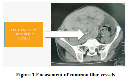
MRI report suggestive of retroperitoneal mass with right iliac blood vessels involvement with right ureter compression causing right hydroureteronephrosis.
Pre-OP HPE report: Malignant fibrous histiocytoma. Before proceeding to surgery, right ureteric stent placement was done (Figure 1).
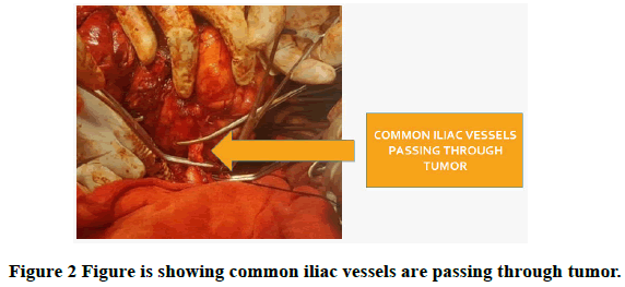
Intra-OP findings: Retroperitoneal mass with right iliac vessels encasement.
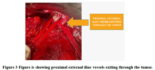
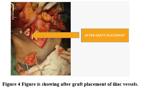
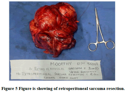
20*15 cm tumor involving right iliac fossa proceeded with mobilisation of terminal ileum and ileocaecal junction, right ureter traced and isolated tumor removed into along with the proximal common iliac and distal external and internal iliac vessels, cut ends of common iliac and external iliac vessel are occluded with vascular clamps. Dacron graft was used to reconstruct the vessels with 5-0 prolene.
Procedure done: Wide local excisions of tumor with right ileofemoral bypass graft.
Post OP HPE report: Malignant fibrosarcoma.
It shows Figures 2-5.
Results and Discussion
Primitive retroperitoneal tumors generate a special interest due to the complexity of the surgical treatment and the high rate of recurrence after a radical operation that constitutes a frequent cause of death [7]. Surgical strategy is dependent on acquiring detailed imaging data in several planes and on 3D reconstructions, as accurate as possible, to gain insight into tumor vascular and visceral relationships. The final diagnosis of primitive retroperitoneal tumor, as well as of histopathologic type, can be achieved only by surgical intervention with the complete examination of the tumor resection piece.
In the past, vascular involvement by the tumor was considered one of the contraindications of surgical resection. Vascular involvement by direct invasion could be seen as an indirect sign of biological aggressiveness. In some cases, the tumor only pushes against major vessels and resecting them only to obtain clear margins is probably a risk that exceeds the potential benefits. There is no data that an extensive vascular resection would increase the local control and ultimately, the patient’s survival, thus the decision for the resection of an adjacent main vessel should be made when the dissection of the vessel is not possible, because of its infiltration or when their dissection would have risks. Venous blood vessels are the most common encased or invaded by the retroperitoneal masses [8,9].
Schwarzbach, et al., have reported in their study that, out of the 25 patients with vascular involvement by retroperitoneal sarcomas, 64% have had venous involvement, 16% of the patients had arterial and venous involvement and arterial-only involvement was observed in 20% of the cases, tumors originating from the vessel walls were less common (8 patients), compared to those that were secondarily invaded (17 patients). In their paper, 22 patients have had vascular reconstruction.
In many centres, now, it is already accepted that in case of vascular involvement by retroperitoneal tumors, ample multiple vascular sacrifices followed by complex arterial and venous reconstructions must be done [10]. 5 years survival rate for all operated patients (radically and unradically) was low, of 14% at 5 years, at the lower limit of the interval reported in specialized literature. No significant difference between the survival rates for the patients with benign or malignant tumors has been noticed, because the resectability of these tumors has not been different. The only factor that has fundamentally improved survival rate has been represented by radical resection, even with the price of visceral sacrifices. Vascular involvement, impeding the achievement of a radical intervention, represented a predictor of a shorter survival of these patients.
Conclusion
Primitive retroperitoneal tumors constitute a very difficult area of general surgery, associating a poor prognosis with high recurrence rates and low disease free survival rates. An adequately trained, multidisciplinary surgical team, including a vascular surgeon, with an appropriate material and equipment basis, experienced in retroperitoneal tumor surgery, can successfully handle ample, high complexity operations and can solve possible ensuing complications associated to such radical, even aggressive interventions.
References
- Pipitone N, Vaglio A and Salvarani C. Retroperitoneal fibrosis. Best Practice and Research Clinical Rheumatology, Vol. 26, No. 4, 2012, pp. 439-448.
[Crossref] [Google Scholar] [PubMed]
- Patil SS, Shah VR and Choudhary VA. Rare case of retroperitoneal lipomatosis in an Indian woman. Journal of Radiology Case Reports, Vol. 10, No. 4, 2016, pp. 23-30.
[Crossref] [Google Scholar] [PubMed]
- Wang CY, et al. Extremely rare case of retroperitoneal “ancient” schwannoma with significant cystic change embedded in the psoas muscle. The Kaohsiung Journal of Medical Sciences, Vol. 37, No. 7, 2021, pp. 632-633.
[Crossref] [Google Scholar] [PubMed]
- Levin DC, Watson RC, Baltaxe HA. Arteriography of retroperitoneal masses. Radiology, Vol. 108, No. 3, 1973, pp. 543-551.
[Crossref] [Google Scholar] [PubMed]
- Latona JA, et al. Modified Appleby procedure with arterial reconstruction for locally advanced pancreatic adenocarcinoma: A literature review and report of three unusual cases. Journal of Gastrointestinal Surgery, Vol. 20, No. 2, 2016, pp. 300-306.
[Crossref] [Google Scholar] [PubMed]
- Balaji R, Ramachandran K and Somanathan T. A rare case of solitary fibrous tumour of the sigmoid mesocolon: Imaging features and review of literature. Cancer Imaging, Vol. 9, No. 1, 2009, pp. 67.
[Crossref] [Google Scholar] [PubMed]
- Pace C, Ward S. A rare case of IgG4 related sclerosing disease of the maxillary sinus associated with bone destruction. Journal of Oral and Maxillofacial Surgery, Vol. 68, No. 10, 2010, pp. 2591-2593.
[Crossref] [Google Scholar] [PubMed]
- Kadam SS, et al. Functional malignant retroperitoneal paraganglioma with liver metastasis: A rare case report. Indian Journal of Surgical Oncology, Vol. 12, 2021, pp. 186-192.
[Crossref] [Google Scholar] [PubMed]
- Laroia ST, et al. Malignant retroperitoneal extra gastrointestinal stromal tumor: A unique entity. World Journal of Oncology, Vol. 7, No. 2-3, 2016, pp. 45.
[Crossref] [Google Scholar] [PubMed]
- Katz MH, et al. Retroperitoneal dissection in patients with borderline resectable pancreatic cancer: Operative principles and techniques. Journal of the American College of Surgeons, Vol. 215, No. 2, 2012, pp. 11.
[Crossref] [Google Scholar] [PubMed]

