Research Article - International Journal of Medical Research & Health Sciences ( 2023) Volume 12, Issue 5
Clinical Features of Palpable Breast Masses in Patients Presenting to Tertiary Care Hospital in Northern India: A Prospective Observational Study
Zaheer Ahmed* and Barinder KumarZaheer Ahmed, Department of Surgery, Government Medical College, Rajouri, Jammu and Kashmir, India, Email: khan.shahbaz78@gmail.com
Received: 20-Jan-2023, Manuscript No. IJMRHS-23-87579; Editor assigned: 23-Jan-2023, Pre QC No. IJMRHS-23-87579 (PQ); Reviewed: 06-Feb-2023, QC No. IJMRHS-23-87579; Revised: 26-Apr-2023, Manuscript No. IJMRHS-23-87579 (R); Published: 12-May-2023
Abstract
Background: Breast problems can present themselves in a number of ways like breast pain, nipple discharge, cystic lesions and more commonly as a lump. A lump in the breast is of great concern to the patients and is also a challenge to the diagnostic intelligence and judgement of the surgeon. Fibroadenoma is the most common benign breast mass, invasive ductal carcinoma is the most common malignancy. Most masses are benign, but breast cancer is the most common cancer and the second leading cause of cancer deaths in women.
Materials and methods: The study was conducted over a period of 1½ years in patients attending the surgical OPD of associated hospital, GMC Rajouri. All patients were subjected to complete clinical examination and evaluation. This involved thorough history taking including personal history, especially with respect to previous breast cancer or biopsies with benign histology, family history of breast or ovarian cancer, abnormalities suspicious of malignancy (e.g. palpable mass, skin retraction, nipple discharge). Clinical examination of breast mass included inspection and palpation.
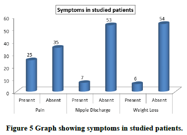
Results: Out of 60 patients, 31 had left sided masses, 28 had right sided masses and 1 had bilateral mass. Most common symptoms of studied patient were pain in 25 followed by nipple discharge in 7 followed by weight loss in 6. Most common site involved was upper outer quadrant, followed by upper inner quadrant, followed by lower outer quadrant, followed by central region followed by lower inner quadrant.
Keywords
Lump, Quadrant, Palpable, Nipple discharge, Ovarian cancer
Introduction
Breast problems can present themselves in a number of ways like breast pain, nipple discharge, cystic lesions and more commonly as a lump. A lump in the breast is of great concern to the patients and is also a challenge to the diagnostic intelligence and judgement of the surgeon. Presence of a three dimensional or space occupying lesion in the breast is the most reliable sign of both benign and malignant disease. However, some non-neoplastic lesions of the breast like traumatic fat necrosis, acute and chronic breast abscess, fibroadenosis, breast cysts, etc. also produce space occupying lumps. Lump in the general sense of the word has a psychological impact on the patient. Of these malignant breast disease is the most dreaded one, feared not only by the patients but also by the surgeons as well. Cases of breast cancer have been recorded in medical writings for more than 5,000 years. In documents from the ancient times, they appear with perhaps greater frequency than any other form of cancer. The first written evidence suggestive of breast cancer is from ancient Egypt. Edwin Smith surgical papyrus, dating back from 3000 to 2500 BC [1,2].
Fibroadenoma is the most common benign breast mass, invasive ductal carcinoma is the most common malignancy [3]. Most masses are benign, but breast cancer is the most common cancer and the second leading cause of cancer deaths in women. Although most breast cancers occur in women older than 50 years, 31 percent of women diagnosed with breast cancer between 1996 and 2000 were younger than 50 years [4].
A complete Clinical Breast Examination (CBE) includes an assessment of both breasts and the chest, axillae and regional lymphatics. In pre-menopausal women, the CBE is best done the week following menses, when breast tissue is least engorged. With the patient in an upright position, the physician visually inspects the breasts, noting asymmetry, nipple discharge, obvious masses and skin changes, such as dimpling, inflammation, rashes and unilateral nipple retraction or inversion. With the patient supine and one arm raised, the physician thoroughly palpates breast tissue on the raised arm side in the superficial, intermediate and deep tissue planes (i.e. the “triple touch” technique), axilla, supraclavicular area, neck and chest wall. Assessing the size, texture and location of any masses. The physician should note the size of the masses to document changes over time. Next, the physician should inspect the areola nipple complex for any discharge. CBE sensitivity can be improved by longer duration (i.e. 5-10 minutes) and increasing precision (i.e. using a systematic pattern, varying palpation pressure, and using three finger pads and circular motions) of palpation [5,6].
Materials and Methods
The present, prospective study was conducted in the postgraduate department of general surgery, government medical college Rajouri.
Inclusion criteria were: All patients with suspicious palpable breast masses.
Exclusion criteria were: All patients with infective masses.
Clinical examination
Clinical examination of breast mass included inspection and palpation. Inspection was done in different postures with:
• Arms by the side of body. In this view general appearance of breast was noted. Any visible swelling, scar,
superficial veins and any other superficial lesions were looked for.
• Arms raised over the head. It was particularly used to look for lumps, skin dimpling, nipple retraction or
deviation of nipples as these are more marked as patient raises her hands above her head.
• Patient sitting and bending forwards so that breasts fall away. It was used to check fixity of breast to chest.
Normal breast falls more than diseased breast suggesting fixity of diseased breast to chest wall or pectoralis
muscle.
• Patient sitting and hands pressed on waist to contract the pectoralis muscle. Skin dimpling becomes more
evident.
• Patient recumbent with 45 degree head end elevation and both hands lying by the side of head, by doing this,
whole breast including the inferior quadrants are seen better.
Palpation of breast was done systematically quadrant wise with the palmer surface of fingers. Palpation was done in sitting posture or patient in recumbent position. Dorsal aspects of fingers were used to ascertain temperature over the breast by comparing diseased breast with normal breast. After this breast tissue was fixed in between fingers of one hand and the lump was pushed to one side with the other hand. If the lump was fixed to breast tissue then breast tissue moved along with lump. This was done to demonstrate fixity of lump to breast tissue. Skin tethering was demonstrated by moving the lump from side to side and observing whether skin dimples appear at extremes of movement. As the malignant lump grows it may infiltrate the cooper’s ligament and make these strands inelastic and pull the skin inwards rendering puckering of skin. Skin fixity was demonstrated by trying to lift the skin from the underlying lump in between fingers. If skin was fixed to the underlying lump, the overlying skin could not be lifted up from the underlying lump. Certain skin changes were made more obvious on palpation like peau-d-orange. Peau-d-orange is obvious on inspection but was demonstrated by squeezing a segment of skin over the breast which showed lymphedema in the skin with prominent hair follicles in between. Clinical examination was concluded by examination of axillary lymph nodes. Examination was done by placing patient in sitting posture facing clinician and proper exposure was done. The anterior, central, apical and the lateral group of lymph nodes were palpated from the front and the posterior or subscapular group were palpated from the back of patient. Right axilla was palpated with the left hand except the lateral and posterior group which were palpated by corresponding hand. The results of the observations at the end of the study were entered in Microsoft Excel and descriptive analysis of the data was done. Categorical variables were summarized as frequency and percentage.
Results and Discussion
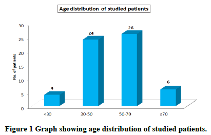
Most common age group affected was 50 to 70 years with a mean + SD of 50.9 ± 14.47. Youngest patient was 26 years old and oldest patient was 81 years old (Table 1 and Figure 1).
| Age (years) | No. of patients | Percentage | Mean ± SD | Range |
|---|---|---|---|---|
| <30 | 4 | 6.7 | 50.9 ± 14.47 | 26-81 |
| 30-50 | 24 | 40.0 | ||
| 50-70 | 26 | 43.3 | ||
| ≥ 70 | 6 | 10.0 | ||
| Total | 60 | 100 |
Table 1. Age distribution of studied patients.
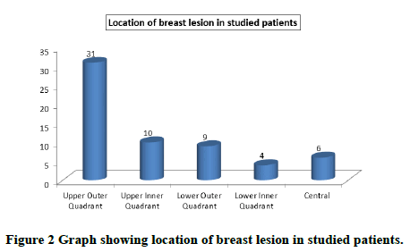
Most common location of breast lesions in patients was upper outer quadrant 31 (51.7%) and least common location was lower inner quadrant 4 (6.6%) (Table 2 and Figure 2).
| Location | No. of patients | Percentage |
|---|---|---|
| Upper outer quadrant | 31 | 51.7 |
| Upper inner quadrant | 10 | 16.7 |
| Lower outer quadrant | 9 | 15.0 |
| Lower inner quadrant | 4 | 6.6 |
| Central | 6 | 10.0 |
Table 2. Location of breast lesion in studied patients.
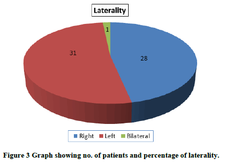
Left sided lesions 31 (51.67%) were more than right sided lesions 28 (46.67%) with only 1 (1.66%) bilateral lesion (Table 3 and Figure 3).
| Laterality | No. of patients | Percentage |
|---|---|---|
| Right | 28 | 46.67 |
| Left | 31 | 51.67 |
| Bilateral | 1 | 1.66 |
Table 3. No. of patients and percentage of laterality.
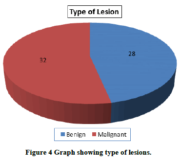
Benign lesions were present in 28 (46.33%) patients and malignant lesions were present in 32 (53.33%) patients (Table 4 and Figure 4).
| Type of lesions | Number of patients | Percentage |
|---|---|---|
| Benign | 28 | 46.67 |
| Malignant | 32 | 53.33 |
Table 4. Type of lesions.
Pain was the most common symptom present in the patients 25 (41.7%) followed by nipple discharge 7 (11.7%) and then weight loss, which was present in 6 (10%) patients (Table 5 and Figure 5).
| Symptoms | No. of patients | Percentage | |
|---|---|---|---|
| Pain | Present | 25 | 41.7 |
| Absent | 35 | 58.3 | |
| Nipple discharge | Present | 7 | 11.7 |
| Absent | 53 | 88.3 | |
| Weight loss | Present | 6 | 10 |
| Absent | 54 | 90 | |
Table 5. Symptoms in studied patients.
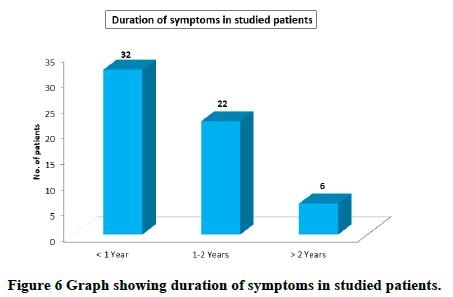
Most patients had duration of symptoms for less than one year; mean duration of symptoms was 1.1 ± 0.93 years. Range was from 1 month to 4 years (Table 6 and Figure 6).
| Duration | No. of patients | Percentage | Mean ± SD | Range |
|---|---|---|---|---|
| <1 year | 32 | 53.3 | 1.1 ± 0.93 | 1 month to 4 years |
| 1-2 years | 22 | 36.7 | ||
| >2 years | 6 | 10.0 | ||
| Total | 60 | 100 |
Table 6. Duration of symptoms in studied patients.
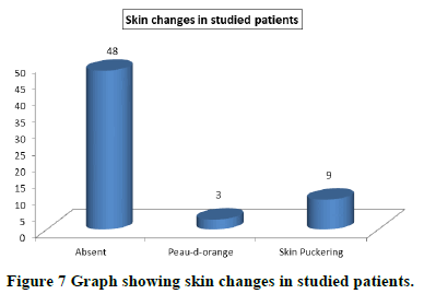
Skin changes were present in 20% patients. Most common skin change was skin puckering which was seen in 9 (15%) patients, peau-d-orange was present in 3 (5%) patients (Table 7 and Figure 7).
| Skin changes | No. of patients | Percentage |
|---|---|---|
| Absent | 48 | 80.0 |
| Peau-d-orange | 3 | 5 |
| Skin puckering | 9 | 15 |
Table 7. Skin changes in studied patients.
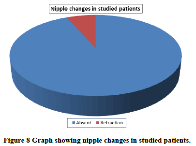
Out of a total of 60 patients, 4 (6.67%) had nipple retraction (Table 8 and Figure 8).
| Nipple changes | No. of patients | Percentage |
|---|---|---|
| Absent | 56 | 93.33 |
| Retraction | 4 | 6.67 |
Table 8. Nipple changes in studied patients.
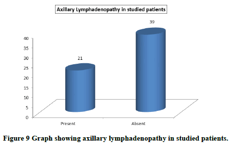
Out of a total of 60 patients, enlarged axillary lymph nodes were seen in 21 (35%) patients (Table 9 and Figure 9).
| Axillary lymphadenopathy | No. of patients | Percentage |
|---|---|---|
| Present | 21 | 35 |
| Absent | 39 | 65 |
Table 9. Axillary lymphadenopathy in studied patients.
Clinical assessment is the most important step and perhaps stepping stone in the diagnosis of any disease which includes breast diseases particularly breast lumps. When a proper clinical assessment is completed even by most experienced clinician, doubt about the diagnosis may still be present, which means that further diagnostic assistance would be needed to come to a most accurate diagnosis. Clinical breast examination is secondary preventive measure that aids in early detection of breast cancer and better management. For average risk asymptomatic women in their 20’s and 30’s, it is recommended that CBE be part of a periodic health examination, preferably at least every three years [7].
Age distribution of the studied patients
Most common age group suffering from the disease in our study was 50-70 years followed by 30-50 with mean age 50.9 years (range 26-81 years). This is in accordance to other reports published from Indian subcontinent which show that carcinoma breast in Indian population occurs a decade earlier than western group [8,9]. Almost similar observations were made by Vinod Raina, et al. with mean age of disease 47 years (range 23-82 years) [10].
Location of breast masses
Most common location of mass was upper outer quadrant (51.6%) followed by upper inner quadrant (16.6%), lower outer quadrant (15%), central (10%) and last was lower inner quadrant (6.6%). Similar findings were recorded by Seth Rummel, et al. who reported with following frequency upper outer quadrant (51.5%) was significantly higher than in the upper inner quadrant (15.6%), lower outer quadrant (14.2%), central (10.6%) and lower inner quadrant (8.1%) [11].
Side determination of the lesions
Of the 60 masses 31 involved left breast that is 51.6%, 28 masses involved right breast that is 46.6% and one patient had bilateral mass that is 1.6%. Similar observations were made by Muhammad Naeem, et al. [12]. The disease was found on the left side in (52.1%), on the right side in (43.4%) and there were (4.3%) of bilateral breast masses.
Incidence of benign and malignant breast masses
It was observed that out of 60 cases, there were 28 (46.67%) benign and 32 (53.33%) malignant lesions. This did not correlate well with the study of Todd R Kumm, et al. who reported an incidence of 72% for benign and 28% for malignant lesions [13]. This higher incidence of malignant lesions in our study was possibly because most of benign lesions were uncomplicated and were not included in the study and therefore, were not subjected to triple assessment or MRI and were treated on OPD basis.
Symptoms of the studied patients
Mastalgia: It was present in 41.7% patients. Similar observations were made by Tibor Decholnoky, who noted pain in 33% of his patients. Mastalgia was most common in the age group 26 to 50 years. Cyclical mastalgia was present in 66.6% and non-cyclical mastalgia was present in 33.3% of the patients complaining mastalgia. These results are comparable with the study by SK Katiyar, et al. who found that most frequent age group complaining of mastalgia was 26 to 45 years. They also found that among the patients with mastalgia, cyclical mastalgia was present in 63.44% of the cases whereas non-cyclical mastalgia was present in 36.56% of the cases. Mastalgia was more common in benign masses as compared to malignant masses. Similar results were found by Lumachi, et al. who found mastalgia only in 3.2% patients with malignant masses.
Nipple discharge: It was present in 11.7% patients. Almost similar results were seen by Geller, et al., who reported nipple discharge in 7.6% patients.
Conclusions
The majority of our patients were between 30-70 years. With mean age 50.9 + 14.47 (range 26 -81 years).
• Out of 60 patients who were included, 32 (53.33%) were malignant and 28 (46.67%) were benign.
• Out of 60 patients, 31(51.67%) had left sided masses, 28 (46.67%) had right sided masses and 1 (1.66%) had
bilateral mass.
• Most common symptoms of studied patient were pain in 25 (41.7%) followed by nipple discharge in 7 (11.7%)
followed by weight loss in 6 (10%).
• Most common site involved was upper outer quadrant (51.5%), followed by upper inner quadrant (15.6%),
followed by lower outer quadrant (14.2%), followed by central region (10.6%) followed by lower inner quadrant
(8.1%).
• Most common sign in study patients was axillary lymphadenopathy in 21 (35%) patients followed by skin
changes 12 (20%) patients followed by nipple retraction 4 (6.67%) patients.
Conflicts of Interest
None
References
- Iglehart JD and Kaelin CM. Diseases of the breast. In: Sabiston text book of surgery. 17th edition, Akshantala Enterprises Private Limited, Karnataka, India, 2004;877.
- Schoonjans JM and Brem RF. Fourteen gauge ultrasonographically guided large core needle biopsy of breast masses. Journal of Ultrasound in Medicine, Vol. 20, No. 9, 2001, pp. 967-972.
[Crossref] [Google Scholar] [PubMed]
- Klein S. Evaluation of palpable breast masses. American Family Physician, Vol. 71, No. 9, 2005, pp. 1731-1738.
[Google Scholar] [PubMed]
- Barton M, et al. The rational clinical examination. The screening clinical breast examination: Should it be done? How? JAMA, Vol. 282, No. 13, 1999, pp. 1270-1280.
[Crossref] [Google Scholar] [PubMed]
- Campbell HS, et al. Improving physicians and nurses clinical breast examination: A randomized controlled trial. American Journal of Preventive Medicine, Vol. 7, No. 1, 1991, pp. 1-8.
[Crossref] [Google Scholar] [PubMed]
- Smith RA, et al. American cancer society guidelines for breast cancer screening update. CA: A Cancer Journal for Clinicians, Vol. 53, No. 3, 2003, pp. 141-169.
[Crossref] [Google Scholar] [PubMed]
- Sandhu DS, et al. Profile of breast cancer patients at a tertiary care hospital in north India. Indian Journal of Cancer, Vol. 47, No. 1, 2010, pp. 16-22.
[Crossref] [Google Scholar] [PubMed]
- Murthy MS, et al. Changing trends in incidence of breast cancer; Indian scenario. Indian Journal of Cancer, Vol. 46, No. 1, 2009, pp. 73-74.
[Crossref] [Google Scholar] [PubMed]
- Seth R, et al. Tumour location within the breast: Does tumour site have prognostic ability? Cancer, Vol. 9, 2015, pp. 552.
[Crossref] [Google Scholar] [PubMed]
- Todd RK, et al. Elastography for the characterisation of breast lesions: Initial clinical experience. Cancer Control, Vol. 17, No. 3, 2010, pp. 156-161.
[Crossref] [Google Scholar] [PubMed]
- Decholnoky T. Benign tumors of the breast. Archives of Surgery, Vol. 38, No. 1, 1939, pp. 79-98.
- Lumachi F, et al. Prevalence of breast cancer in women with breast complaints. Retrospective analysis in a population of symptomatic patients. Anticancer Research, Vol. 22, No. 6B, 2002, pp. 3777-3780.
[Google Scholar] [PubMed]
- Geller BM and Barlow WE. Use of the American college of radiology BI-RADS to report on the mammographic evaluation of women with signs and symptoms of breast disease. Radiology, Vol. 222, No. 2, 2002, pp. 536-542.
[Crossref] [Google Scholar] [PubMed]

