Research - International Journal of Medical Research & Health Sciences ( 2023) Volume 12, Issue 3
Effects of Circadian Rhythm Disruption on Cognitive Function and Related Expression of DREAM and Hyperphosphorylated Tau Protein in the Rat's Hippocampus
Norhida Ramli*Norhida Ramli, Medical Lecturer of Basic Medical Sciences, University Malaysia Sarawak, Malaysia, Email: n_hida@yahoo.com
Received: 16-Feb-2023, Manuscript No. ijmrhs-23-89467; Editor assigned: 20-Feb-2023, Pre QC No. ijmrhs-23-89467(PQ); Reviewed: 28-Feb-2023, QC No. ijmrhs-23-89467(Q); Revised: 16-Mar-2023, Manuscript No. ijmrhs-23-89467(R); Published: 28-Mar-2023
Abstract
Introduction: Sleep deprivation has been demonstrated to impair the cognitive function associated with Downstream Regulatory Element Antagonistic Modulators (DREAM), Brain-derived neurotrophic factor (BDNF), and cAMP-response element binding (CREB) protein expression changes in rat hippocampus. This study was conducted to investigate whether Circadian rhythm Disruption (CD) has a similar mechanism with sleep deprivation on cognitive function and DREAM, Tau, and hyperphosphorylated Tau protein expression changes. Methods: 24 male Sprague Dawley rats were equally divided into 3 groups: (i) control (C), (ii) Acute CD (ACD), and (iii) Chronic CD (CCD). 1 cycle of the CD consists of phase-shifting, where daily light off was phased advanced three hours daily for 6 consecutive days followed by ten days of re-entrainment for recovery. The acute CD was induced by a single cycle. In contrast, the chronic CD was induced by 4 cycles of the CD. The C group was set in regular daily 12 hours light/dark. The rat's spatial learning and memory were measured by the Morris water maze test followed by immunohistochemistry analysis for protein expression. Results: There was no significant difference in escape latency during the acquisition trial and swim time in the target quadrant during the probe test between all groups. The hippocampus DREAM, Tau, and hyperphosphorylated Tau protein expression also were not statistically significant differences compared to all groups. Conclusion: We concluded that in this study, the CD has not adequately elicited changes in cognitive function and expression of the hippocampus DREAM, Tau, and hyperphosphorylated Tau proteins.
Keywords
Circadian rhythm, DREAM protein, Tau protein, Morris Water Maze test, Hippocampus
Introduction
The physiological alteration response to light and darkness in a living organism is called the circadian rhythm. For instance, the body temperature indicates the basal metabolic activities are higher in the evening before our sleeping time and back to the lowest point early in the morning. We felt sleepy at night and awake during the light day symbolizes a normal circadian rhythm regulation of the sleep-wake cycle [1]. Circadian rhythms are found throughout all phylogenies that include animal, plant, and microscopic organisms. The circadian rhythms regulate various physiological events that include the cell cycle, body temperature, metabolism, feeding, and the sleep-wake process [2].
Modern society is now becoming less dependent on the natural 24-hour light-dark cycle. The need to fulfill the demand for excellent job performance, high academic achievement, and a work shift implementation worsened the situation. All of these scenarios make our lifestyle in this contemporary era increasingly stressful. Failure to manage it may be affecting the quality and quantity of our sleep needed and disrupt our normal circadian rhythm. The endogenous circadian clock and the sleep-wake cycle misalignment can produce a "jet lag disorder" associated with fatigue symptoms, gastrointestinal distress, reduced psychomotor coordination, and reduced cognition and mood [3]. Industrial workers and nurses exposed to frequent shift work have been reported to produce cognitive performance deficits [4, 5]. Therefore, the relationship between circadian rhythm, sleep, and cognitive function has broad implications for public health.
The formation of new memories involves changes in synaptic efficacy and membrane excitability at the hippocampus's cellular level [6]. Some suggest that these neuronal properties can be altered during periods of sleep and by sleep disruption [7]. Indeed, a study has shown that Rapid Eye Movement (REM) sleep deprivation can alter neuronal excitability and reduce the strength of long-term potentiation, a cellular marker of memory, and modifies the cellular composition of the sub-granular zone [8]. Previous studies in our lab have shown that REM sleep deprivation impairs learning and memory processes by modulating Downstream Regulatory Element Antagonistic Modulators (DREAM), cAMP-Response Element-Binding (CREB), and Brain-Derived Neurotrophic Factor (BDNF) proteins expression in the rat hippocampus [9, 10].
Learning and memory processes have been found to involve DREAM protein modulation [11]. DREAM protein acts as a negative regulator of the transcription factor for memory, CREB, in a Ca2+ -dependent manner [12]. A knockout DREAM gene study has been shown markedly enhances cognitive function by facilitating CREBdependent transcription. DREAM protein also has been found to act as a negative regulator of the NMDA receptor function in hippocampal synaptic plasticity [13]. Therefore, modulation of DREAM protein in hippocampal areas is suggested to be involved in the mechanisms of REM sleep deprivation-induced learning and memory impairment in rats .
The microtubule-associated protein Tau is crucial in stabilizing neurofilaments in neurons [14]. Studies have reported that Tau protein is hyperphosphorylated and aggregates to form intracellular neurofibrillary tangles in neurons and affects cognitive functions after REM sleep deprivation in mice [15, 16]. Thus, it can be suggested that REM sleep deprivation can induce increases in DREAM and hyperphosphorylated Tau protein expression in the rodent's brain and affect cognitive function. Human subjects who could not have proper sleep duration or poor sleep quality, such as sleep apnea patients, also tend to have poor memory and other cognitive function deficits. Sleep deprivation is also linked with Alzheimer's Disease (AD), which is associated with a decline in the hippocampus's neurons than a healthy person [17].
In the brain suprachiasmatic nucleus cells, the dark/light cycle is regulated by kinase responses, ultimately by transcription factor CREB [18]. CREB has also been involved in learning and memory processes by regulating DREAM protein (9-10) and Tau protein transcription [19]. However, whether the CD effect on learning and memory processes occurs in similar passions like REM sleep deprivation that affected those protein expressions is still unknown. This study suggested that the CD does not impair spatial learning and memory behavior associated with no changes in DREAM, Tau, and hyperphosphorylated Tau proteins expression in the hippocampus, not similar to REM sleep deprivation.
Method
Animal’s Preparation
We used 24 male Sprague Dawley rats in this study. They were purchased from the Animal Research and Service Centre, Universiti Sains Malaysia (ARASC, USM) at 8 weeks to 10 weeks old. They were acclimatized one week before experiments by daily handling. During acclimatization, they were house paired in a propylene cage to avoid social isolation under the 12:12 light: dark cycle and given water and food ad libitum. The rats were divided equally into three groups: acutely CD rats (Acute; n=8), chronically CD (Chronic; n=8), and control rats (control; n=8). All the experimental animal procedures in this study were approved by the Animal Ethics Committee of Universiti Sains Malaysia (AECUSM).
Disruption of Circadian Rhythm
We selected Craig and McDonald's (2008) protocol to induce CD [20, 21]. Both groups of CD rats were housed in pairs in polypropylene cages with food and water ad libitum. Protocol for CD includes a single cycle for a phaseshifting experiment, consisting of 6 days of phase-shifting followed by 10 days of re-entrainment for recovery. In these 6 days of the phase-shifting stage, daily light off was phased advanced three hours daily for six consecutive days (9:12 hours light/dark cycle). Subsequently, on the 7th through the 16th day, the rats were allowed for reentrainment for recovery. During this re-entrainment phase, the light/dark cycle was put back at a normal level for 12:12 hours (Table 1).
| Days | Light on at | Lights off at |
|---|---|---|
| 1 | 07:30 | 16:30 |
| 2 | 04:30 | 13:30 |
| 3 | 01:30 | 10:30 |
| 4 | 22:30 | 07:30 |
| 5 | 19:30 | 04:30 |
| 6 | 16:30 | 01:30 |
| 7-6 | 13:30 | 22:30 |
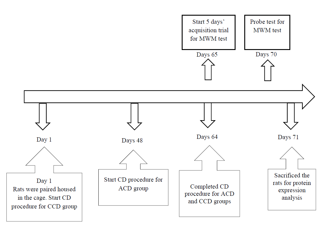
The acute phase-shifting group was put in a single cycle of the phase-shifting stage. Meanwhile, the chronic phaseshifting group was arranged in four cycles of the phase-shifting stage. At the end of the process, all rats were monitored for any weight changes. As for rats in the control group, they were paired house in standard polypropylene cages with food and water ad libitum, on a 12:12 hours light/dark cycle with lights off at 7:30 pm and lights on at 7: 30 am. The procedure of the experiment was showed in Figure 1. NAFLD- Patients with USG evidence of fatty changes in the liver.
Immunohistochemistry (IHC) Analysis
Rats were sacrificed by an intraperitoneal overdose injection of sodium pentobarbitone (100 mg/kg) for IHC analysis after 24 hours Morris Water Maze (MWM) test. The trans cardinal perfusion-fixation method was performed by perfusing 200 ml of Phosphate-Buffered Saline (PBS), followed by 500 ml of ice-cold, 4% paraformaldehyde in 0.1 M Phosphate Buffer (PB) (pH=7.4) to fix the entire organ. The brain was dissected and immediately post-fixed in 4% paraformaldehyde for 4 hours at 4°C, and then cry-protected in 20% sucrose overnight at 4°C. The IHC staining procedure was followed in our previous studies [9]. The brain was embedded in a tissue-freezing medium and sectioned with cryostat microtome at 30 μm sections. The brain section was collected to a 24-well plate as a free-floating section in PBS. Then, the brain sections were incubated overnight with rabbit polyclonal primary antibodies for DREAM PA5-30605 (dilution 1:500), rabbit polyclonal antibody Tau PA114423 (dilution 1:1000), and phosphor-PHF-tau pSer202/Thr205 monoclonal antibody (AT8) MN1020 (dilution 1:800) (Thermo Fisher Scientific, USA). The brain sections were then incubated with biotinylated secondary antibody anti-rabbit (dilution 1:200) for 1 hour.
After 1 hour, the brain sections were reacted with Avidin-Biotin Complex (ABC) and stained with diaminobenzidine and hydrogen peroxide until brown coloration was seen. Sections were examined using an image analyzer (LeicaMPS 60) at ×4 and ×10 magnifications. 4 sections were randomly taken from 1 rat in each group. Three areas of view in the CA region (CA, CA2, and CA3) and one area in the hippocampus DG were chosen from one section. The DREAM positive neurons (DPN), Tau Positive Neurons (TPN), and phosphorylated Tau Positive Neurons (PTN) were confirmed and counted as a mean total DPN, TPN, and PTN for all hippocampus areas for each section. Two blinded investigators have done this procedure. Tau protein that was hyperphosphorylated was measured by the ratio of total PTN over total TPN. The total PTN and TPN were derived from all hippocampus areas that are co-localized in a similar section.
Statistical Analysis
Statistical analyses were performed using the Statistical Package of Social Sciences (SPSS) software version 21. Escape latency was analyzed using repeated measurements ANOVA, and differences between specific groups were analyzed by Bonferroni post hoc test. The differences between groups for swim time spent in the target quadrant, DPN, TPN, and PTN expression were measured using one-way ANOVA, Bonferroni, and Dunnet's post hoc test. All data are reported as mean ± Standard Error Mean (SEM), and the level of significance was set at p<0.05.
Results
Escape Latency (EL)
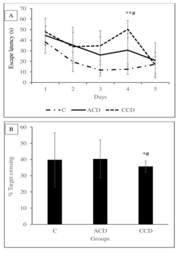
The analysis was done within the daily score in all groups and between the groups' differences for escape latency using the 1-way repeated measure ANOVA test. Generally, it showed no significant reduction in EL time during the 5 days test (p<0.05) within all groups. EL time also was found not to be a significant difference between all groups on each five acquisition days (p>0.05) (Figure 2A).
Probe Test
The probe test was carried out after the completion of five days of spatial learning acquisition. This test aims to assess memory retention. It was done for only one session on the 6th day of the MWM test. The target quadrant, where the platform from previous spatial learning acquisition was located, was removed during this session. During the probe test day, the rat could swim to search the target quadrant's platform for 60 seconds. The duration (in seconds) of visits to the target quadrant was analyzed using the one-way ANOVA test. We found that the time of swimming in the target quadrant was no significant difference between all groups (Figure 2B).
Immunohistochemical Analysis
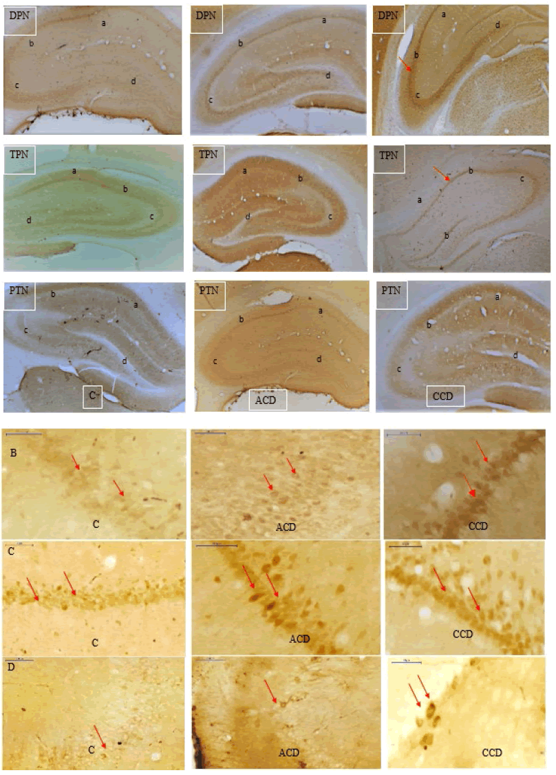
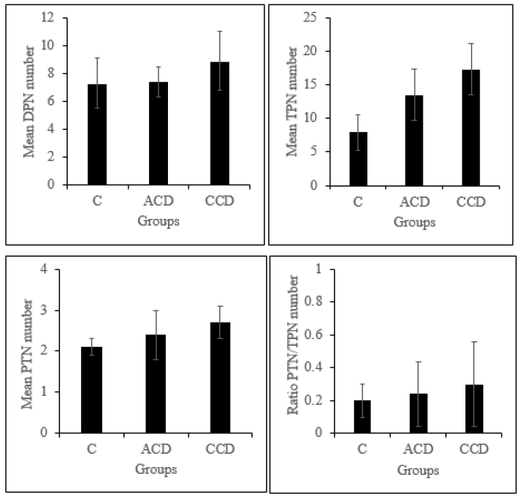
The hippocampus areas for IHC counted were shown in Figure 3A. DPN was found not to be a significant difference between all groups in all areas of the hippocampus (Figure 3B). Statistical analysis also found that the TPN expression was not significantly different from all groups. Similarly, the PTN expression was also found not significantly different between all groups. The hyperphosphorylated tau protein measurement was carried out by calculating the total PTN protein ratio over the total Tau protein expression in the rat brain hippocampus. We found that the ratio was not different between all groups (Figure 3C-3D and Figure 4).
Figure 3. Immunohistochemistry results show the DPN, TPN, PTN (Arrow mark) (4Ã? magnification) at CA1 (a), CA2 (b) CA3 (c), and DG (d) of hippocampus regions of all experimental groups (A), DPN (B), TPN (C) and PTN (D) (Arrow mark) (40Ã? magnification) at random areas of the hippocampus of all experimental groups
Discussion
This study found that the CD does not impair spatial learning and memory with no hippocampus protein expression changes was found. These effects are not similar to the previous study in our lab on REM sleep deprivation and cognitive performance [9, 10]. We cannot see the impairment of spatial learning and memory due to CD in the Morris Water Maze (MWM) test. A rat's ability to acquire a new lesson is tested during the acquisition trial, while retained memory is tested in the probe trial of the MWM test. The escape latency time for C and ACD groups showed a daily reduction, which indicates the learning process is not affected in these groups. However, it was increased in the CCD group, on day 4, but was found no significant difference compared to other groups. The duration to visit the target platform quadrant in the probe test was also found shortest in the CCD group; however not statistically significant compared to the other groups. This effect means that the CCD group only suffered minor memory retention problems. The CCD group was the group that received the chronic phase-shifted light and dark cycle procedure. This finding also was not consistent, which suggested that the chronically phase-shifted light and dark cycle in the rats can affect the learning and memory processes. The possible explanation for these effects is still unclear. It might be due to the different capabilities to release melatonin by the pineal gland and rats' internal physiological regulation to modulate the circadian rhythm changes in the CD experiment in this study [22].
In addition to the behavior finding, we also found no changes in DREAM, Tau, and hyperphosphorylated Tau protein expression in all groups' hippocampus. Thus, the quality of sleep that is not much affected in the CD experiment possibly are the factors that contribute to no changes in spatial learning and memory and those proteins expression in the hippocampus. A few previous studies support the DREAM protein's upregulation in the rats' hippocampal area after sleep deprivation [9, 10]. DREAM protein has been demonstrated in learning and memory by acting as a transcriptional repressor for CREB, a transcription factor in a calcium-dependent manner that involves the learning and memory processes. DREAM gene knockout mice have been reported can facilitate the CREB gene transcription and enhance learning and memory performance [11]. In this study, the CD experiment condition was found to not changes the DREAM protein expression. It is possible that the DREAM protein did not stimulate intracellular calcium that induced a noradrenaline-mediated increase in Na-K-ATPase activity in rat brains [23]. Thus, in this study, the excitatory and inhibitory neurotransmitter release in the hippocampus is not much affected and does not affect brain functionality [24-26]. In this study also, the CD does not show any Tau and hyperphosphorylated Tau protein changes. Under normal physiological regulation of Tau protein, phosphorylation is essential in maintaining microtubule structure in the axon, dendrites, and neurons. Many studies have found an association of hyperphosphorylated Tau proteins with neurodegenerative diseases like Alzheimer's disease and brain injury, either induced by trauma or stress [17]. The excessive accumulation and premature phosphorylation of Tau protein will form the hyperphosphorylated Tau protein, leading to the formation of intracellular neurofibrillary tangles, which affect the normal function of neurons [15, 16]. A study also has demonstrated that the circadian rhythm can be altered at both the behavioral and molecular levels in a mouse model of tauopathy [27]. However, in this study, the CD procedure does not affect the hippocampus's cognitive behavior and protein expression. The explanation for this effect is still unknown, but it suggests that CD-induced memory impairment is a complex phenomenon that is yet to be understood clearly [28].
Conclusion
In conclusion, we suggest that in this study, the CD does not affect the spatial learning and memory function and DREAM, Tau, and hyperphosphorylated Tau proteins expression in the rat's hippocampus. The mechanism for cognitive impairment due to REM sleep deprivation appears not similar to the CD experiment in this study.
Declarations
Conflict of Interest
The authors declared no potential conflicts of interest with respect to the research, authorship, and/or publication of this article.
Acknowledgments
We want to thank the Physiology Laboratory, Cranial, Maxillofacial, and ARASC staff for their technical assistance. We also thanked Malaysia's Government for the Fundamental Research Grant Scheme (FRGS 203/ PPSK/6171130).
References
- Zee, Phyllis C., and Michael V. Vitiello. "Circadian rhythm sleep disorder: irregular sleep wake rhythm." Sleep medicine clinics, Vol. 4, No. 2, 2009, pp. 213-18.
Google Scholar Crossref - Archer, Simon N., et al. "Mistimed sleep disrupts circadian regulation of the human transcriptome." Proceedings of the National Academy of Sciences, Vol. 111, No. 6, 2014, pp. E682-E691.
Google Scholar Crossref - Horsey, Emily A., et al. "Chronic jet lag simulation decreases hippocampal neurogenesis and enhances depressive behaviors and cognitive deficits in adult male rats." Frontiers in Behavioral Neuroscience, Vol. 13, 2020, pp. 1-14.
Google Scholar Crossref - Chellappa, Sarah L., Christopher J. Morris, and Frank AJL Scheer. "Effects of circadian misalignment on cognition in chronic shift workers." Scientific reports, Vol. 9, No. 1, 2019, p. 699.
Google Scholar Crossref - Saleh, Abdelbaset M., et al. "Impacts of nursesâ?? circadian rhythm sleep disorders, fatigue, and depression on medication administration errors." Egyptian Journal of Chest Diseases and Tuberculosis, Vol. 63, No. 1, 2014, pp. 145-153.
Google Scholar Crossref - Abel, Ted, and K. Matthew Lattal. "Molecular mechanisms of memory acquisition, consolidation and retrieval." Current opinion in neurobiology, Vol. 11, No. 2, 2001, pp. 180-87.
Google Scholar Crossref - Prince, Toni-Moi, and Ted Abel. "The impact of sleep loss on hippocampal function." Learning & Memory, Vol. 20, No. 10, 2013, pp. 558-69.
Google Scholar Crossref - Soto-Rodriguez, Sofia, et al. "Rapid eye movement sleep deprivation produces long-term detrimental effects in spatial memory and modifies the cellular composition of the subgranular zone." Frontiers in Cellular Neuroscience, Vol. 10, 2016, pp. 1-13.
Google Scholar Crossref - Abd Rashid, Norlinda, et al. "Acute nicotine treatment attenuates learning and memory impairment in REM sleep deprivation through modulation of CREB and BDNF protein expression in ratâ??s hippocampus." CNS, Vol. 3, No. 2, 2017, pp. 76-84.
Google Scholar - Abd Rashid, Norlinda, et al. "Nicotineâ?prevented learning and memory impairment in REM sleepâ?deprived rat is modulated by DREAM protein in the hippocampus." Brain and behaviour, Vol. 7, No. 6, 2017, p. e00704.
Google Scholar Crossref - Fontan-Lozano, Angela, et al. "Lack of DREAM protein enhances learning and memory and slows brain aging." Current Biology, Vol. 19, No. 1, 2009, pp. 54-60.
Google Scholar Crossref - Naranjo, Jose R., and Britt Mellstrom. "Ca2+-dependent transcriptional control of Ca2+ homeostasis." Journal of Biological Chemistry, Vol. 287, No. 38, 2012, pp. 31674-80.
Google Scholar Crossref - Zhang, Ying, et al. "The DREAM protein negatively regulates the NMDA receptor through interaction with the NR1 subunit." Journal of Neuroscience, Vol. 30, No. 22, 2010, pp. 7575-86.
Google Scholar Crossref - Brandt, Roland, and Lidia Bakota. "Microtubule dynamics and the neurodegenerative triad of Alzheimer's disease: the hidden connection." Journal of neurochemistry, Vol. 143, No. 4, 2017, pp. 409-17.
Google Scholar Crossref - Qiu, Hongyan, et al. "Chronic Sleep Deprivation Exacerbates Learning-Memory Disability and Alzheimerâ??s Disease-Like Pathologies in AβPPswe/PS1Î?E9 Mice." Journal of Alzheimer's Disease, Vol. 50, No. 3, 2016, pp. 669-85.
Google Scholar Crossref - Rothman, Sarah M., et al. "Chronic mild sleep restriction accentuates contextual memory impairments, and accumulations of cortical Aβ and pTau in a mouse model of Alzheimer's disease." Brain research, Vol. 1529, 2013, pp. 200-208.
Google Scholar Crossref - Musiek, Erik S., David D. Xiong, and David M. Holtzman. "Sleep, circadian rhythms, and the pathogenesis of Alzheimer disease." Experimental & molecular medicine, Vol. 47, No. 3, 2015, pp. e148-e48.
Google Scholar Crossref - Hughes, Steven, et al. "Photic regulation of clock systems." Methods in enzymology, Vol. 552, 2015, pp. 125-43.
Google Scholar Crossref - Iqbal, Khalid, et al. "Tau in Alzheimer disease and related tauopathies." Current Alzheimer Research, Vol. 7, No. 8, 2010, pp. 656-64.
Google Scholar Crossref - Craig, Laura A., and Robert J. McDonald. "Chronic disruption of circadian rhythms impairs hippocampal memory in the rat." Brain research bulletin, Vol. 76, No. 1-2, 2008, pp. 141-51.
Google Scholar Crossref - Dubocovich, M. L., et al. "Effect of MT1 melatonin receptor deletion on melatoninâ?mediated phase shift of circadian rhythms in the C57BL/6 mouse." Journal of pineal research, Vol. 39, No. 2, 2005, pp. 113-20.
Google Scholar Crossref - Dubocovich, M. L., et al. "Effect of MT1 melatonin receptor deletion on melatoninâ?mediated phase shift of circadian rhythms in the C57BL/6 mouse." Journal of pineal research, Vol. 39, No. 2, 2005, pp. 113-20.
Google Scholar Crossref - Das, G., et al. "Stimulatory role of calcium in rapid eye movement sleep deprivationâ??induced noradrenaline-mediated increase in Na-K-ATPase activity in rat brain." Neuroscience, Vol. 155, No. 1, 2008, pp. 76-89.
Google Scholar Crossref - Fontanâ?Lozano, Angela, et al. "The Mâ?current inhibitor XE991 decreases the stimulation threshold for longâ?term synaptic plasticity in healthy mice and in models of cognitive disease." Hippocampus, Vol. 21, No. 1, 2011, pp. 22-32.
Google Scholar Crossref - Mohammed, Haitham S., et al. "Neurochemical and electrophysiological changes induced by paradoxical sleep deprivation in rats." Behavioural brain research, Vol. 225, No. 1, 2011, pp. 39-46.
Google Scholar Crossref - Xie, Fang, et al. "Anesthetic propofol normalized the increased release of glutamate and γ-amino butyric acid in hippocampus after paradoxical sleep deprivation in rats." Neurological research, Vol. 37, No. 12, 2015, pp. 1102-07.
Google Scholar Crossref - Stevanovic, Korey, et al. "Disruption of normal circadian clock function in a mouse model of tauopathy." Experimental neurology, Vol. 294, 2017, pp. 58-67.
Google Scholar Crossref - Newman, Abigail W., et al. "Brief circadian rhythm disruption does not impair hippocampal dependent memory when rats are over-trained and given more re-entrainment days." Learning and Motivation, Vol. 69, 2020.
Google Scholar Crossref


