Research - International Journal of Medical Research & Health Sciences ( 2021) Volume 10, Issue 1
Prevalence of Anti-nuclear Antibody Pattern-one Year Experience in a Tertiary Care Center
Reshmy GS*, Mrudula EV, Sumitha Prabhu PS, Dr Ashika MS, Dr Sumithra N Unni and Dr Sajitha Krishnan PReshmy GS, Department of Biochemistry, Amrita Institute of Medical sciences and Research Centre, Amrita Vishwa Vidyapeetham, Kochi, Kerala, India, Email: reshmygs@aims.amrita.edu
Received: 02-Dec-2020 Accepted Date: Jan 22, 2021 ; Published: 29-Jan-2021
Abstract
Autoimmunity is the failure of an organism to recognize its own constituent part as self, thereby allowing immune response against its own cells or tissues. ANA is a marker of the autoimmune process. It is positive for a variety of autoimmune diseases. The study was trying to find the prevalence of ANA in correlation with sex, age group and pattern of ANA .Current study findings show that women and elderly people are more prone to develop autoimmune disorders. The female dominance in ANA production suggests that hormonal or other factors in women play a role in this process. The intrinsic variations in the aging of the immune or endocrine systems may be the reason for ANA to be common in elderly people. Our study showed speckled pattern as the most prevalent pattern, this can be because of its less specificity .All this above mentioned criteria’s should be considered while evaluating a positive ANA result.
Keywords
ANA (Anti-Nuclear Antibody), Female dominance, Speckled pattern, Elderly people
Introduction
Autoimmunity is the failure of an organism to recognize its own constituent part as self, thereby allowing immune response against its own cells or tissues. The exact cause of autoimmune disorders is not yet known. However, this condition may arise due to genetic disorders, certain bacteria and viruses that affect the T-suppressor cell system, medications that affect T-suppressor cells, sequestrated antigens produced within the body etc. Anti-Nuclear Antibodies (ANAs) are unusual antibodies detectable in the blood that have the capability of binding to certain structures within the nucleus of the cells. ANA have been categorized in to 2 main groups. Which include antibodies against single and double-stranded DNA (dsDNA) and histones, and auto antibodies that target other nuclear antigens named as ENA (Extractable Nuclear Antigen) as originally they were extracted from the nuclei with saline? ENA further sub classified as Ribonucleoproteins (RNP), SSA/Ro, or SSB/La, Scl-70, Jo-1, Sm [1].
There are over eighty auto immune disorders known each with its own unique symptoms. But autoimmune disease do share some common characteristic symptoms such as extreme fatigue, joint pain, muscle weakness, swollen glands, inflammation, susceptibility to infections, sleep disturbances, weight loss or gain, low blood sugar, blood pressure change, allergies, digestive problems, anxiety or depression, memory problems, recurrent headaches, low grade fever, recurrent miscarriages etc. The diagnosis of an autoimmune disease is based upon signs, symptoms and laboratory test results.
ANA is a marker of the autoimmune process. It is positive for a variety of autoimmune diseases but is not specific. Consequently, if ANA is positive it is often followed up with other tests associated with arthritis and inflammation such as Rheumatoid Factor (RF), Erythrocyte Sedimentation Rate (ESR), C-Reactive Protein (CRP) and /or complement proteins.
Techniques for ANA Detection
Presence of auto antibodies in the sera of the patient constitutes one of the criteria used for diagnosis of Connective Tissue Disorder (CTD) besides clinical diagnosis the ANA sub typing also helps in identifying a specific CTD. Al-though a battery of laboratory tests are available for ANA detection indirect Immunofluorescence Anti-nuclear Antibody test (IF-ANA) and Enzyme Immunoassay (EIA)/Enzyme Linked Immunosorbent Assay (ELISA) are commonly used in day to day practice.
Anti-Nuclear Antibody-Immuno Fluorescence Assay (ANA-IFA): The Standard ANA Testing Technique
Before development of ANA IFAtest, LE (Lupus Erythematosus) cell preparation was the only method used for diagnosis of SLE (Systemic Lupus Erthematosus). IFA-ANA was designed by George Friou in 1957 since then it has been the most widely used test for diagnosis of CTD. It is inexpensive and easy to perform, with high sensitivity and specificity. The test detects the presence of ANA in the blood of the patient which adheres to reagent test cells (substrate), forming distinct fluorescence patterns that are associated with certain autoimmune diseases. Initially different substrates like tissue sections, desquamated cells, chicken erythrocytes and HeLa cells were tried but later on tissue sections using rat liver or a composite multi block substrate (mouse stomach, rat liver and kidney) became the standard substrate. In 1975 HEp-2 cells were introduced which have further increased the sensitivity of the test. These are the cultured cells of laryngeal squalors cell carcinoma and are available commercially in the form of prefixed on glass slides. Majority of the laboratories around the world are now using HEp-2 cell substrates.
The correct interpretation of the IF-ANA results is important and must always be correlated with the patient’s symptoms and signs. While reporting IF-ANA three parameters are evaluated; these include the pattern of fluorescence, substrate used and the titer of a positive test. A negative IF-ANA result essentially excludes possibility of active CTD.
Different staining patterns are reported which give clues as to the significance of the ANA and type of CTD.
Homogenous (Diffuse): The resting cells have fairly uniform staining. The dividing cells have intense, uniform staining of the chromatin. RIM (Peripheral): It is a type of homogenous pattern where staining is more at the periphery of the nucleus and so appear as a rim.
Speckled
Fine Speckled: Numerous small uniform points of fluorescence scattered throughout the nucleus with distinct nuclear margins. The dividing cell chromatin negative.
Coarse speckled: Medium sized uniform points of fluorescence scattered throughout the nucleus with distinct nuclear margins. Here also dividing cell chromatin is negative.
Centromere
The resting cell nuclei have numerous discrete speckles. If it were possible to count them most cells would have 46 dots, same as the number of chromosomes present in human cells. The chromatin of the dividing cells has a zip like staining pattern
Nucleolar
Nuclei of resting cells have staining of the nucleolar regions. The dividing cell chromatin may be negative, positive, or stain with a few dots
Objectives
The study was an attempt to find the prevalence of ANA in correlation with sex, age group and pattern of ANA.
Materials and Methods
The study was carried out at Amrita Institute of medical Sciences and Research centre, Kerala, India during the period of January 2018 to December 2019, a one year experience. ANA-IFA was done in 3312 samples using immunofluorescence method in Bio-Rad Phd.
The staining pattern is determined and recorded. The intensity is graded using the following criteria
4+ Brilliant apple green fluorescence
3+ Bright apple green fluorescence
2+ Clearly distinguishable, positive fluorescence
1+ lowest specific fluorescence
Results
In this retrospective study of one year time period, a total of 3312 samples were analyzed, of these 902 samples were positive for ANA Screening by immunofluorescence.
Mainly four types of patterns were obtained for ANA IFA.
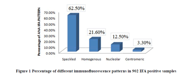
The Speckled Pattern is the most common pattern observed 564/902 (62.5%), followed by Homogenous pattern 195/902 (21.6%), nucleolar pattern 113/902 (12.5%) and centromeric pattern 30/902 (3.3%). Most prevalent pattern obtained by the study was speckled pattern (Table 1 and Figure 1).
| IFA Pattern | Number | Percentage |
|---|---|---|
| Speckled | 564 | 62.50% |
| Homogenous | 195 | 21.60% |
| Nucleolar | 113 | 12.50% |
| Centromeric | 30 | 3.30% |
Table 1: Percentage of different immunofluorescence patterns in 902 IFA positive samples
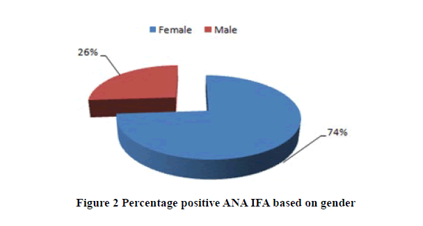
Out of the 902 positive samples 666 samples were females and 236 samples were male’s .The most popular gender with positive ANA according to the current study was females (Table 2 and Figure 2).
| Gender | Percentage |
|---|---|
| Females | 73.80% |
| Males | 26.10% |
Table 2: Percentage positive ANA IFA based on gender
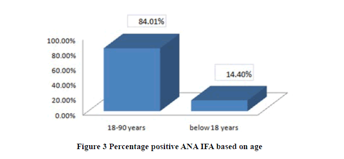
Study was also focusing on the age of the patients and found that elderly people are more prone to autoimmune diseases. Below 18 years there were only 130 patients positive for ANA IFA and between 18-90 years it was 758 patients (Table 3 and Figure 3).
| Age | Percentage |
|---|---|
| 18-90 years | 84.01% |
| Below 18 years | 14.40% |
Table 3: Percentage positive ANA IFA based on age
Discussion
One of the most common hallmarks of autoimmune disorders is the presence of various auto antibodies in the sera of patients affected by these disorders. In the late 1950’s a novel technique that utilized indirect immunofluorescence was developed [2]. This was found to provide a more sensitive assay for the detection of the autoimmune phenomenon. The IFA is considered as the “gold standard” for testing ANA in clinical practice. By using Hep-2 cells as substrates, the IFA permits the detection of antibodies to more than 30 different nuclear and cytoplasmic antigens comprising more than 50 auto antibodies [3].
ANA IFA results are usually reported based on 4 basic patterns Homogenous, Speckled, Nucleolar, Centromeric. In addition titers are usually reported for each pattern seen in ANA IFA testing [3]. Though positive fluorescent staining indicates the presence of ANA, it doesn’t, however allow precise identification of these antibodies. For this purpose; specialized techniques such as ELISA, western blotting or line immunoassay are employed.
The study was an attempt to find the most prevalent gender, age group and type of ANA IFA pattern. Out of the 902 positive ANA IFA samples, 666 were females and 236 were males. 758 of the patients were between the age group of 18-90 years and only 130 were below 18 years. These findings show that females and elderly people are more prone to develop autoimmune disorders. The findings of the present study are similar to the work done by Satoh M et al, which points out that 32 million persons in the US have ANA and the prevalence is higher among females, older individuals, African Americans and those with a normal body weight [4]. The reason for the female predominance in autoimmune diseases is not completely understood. The female dominance in ANA production suggests that hormonal or other factors in females play a role in this process [4]. Estrogen affects the immune system because all immune cells have estrogen receptors and these hormones encourage the immune cells to begin to make too many antibodies. Symptoms of autoimmune diseases get worsened when estrogen level increase for example, during the menstrual cycle and during pregnancy. Since women have a more vigorous immune response due to estrogen their immune systems might be more likely to become hyperactive.
Our reports suggest that aging is associated with autoimmunity and the prevalence of ANA is higher in the elderly. This may be because of the intrinsic variations in the aging of the immune or endocrine systems or sampling bias. A similar observation was given by Nilsson B O that increased prevalence of ANA is seen in the oldest-old. He also emphasized the importance of taking gender and age into consideration when evaluating ANA [5].
Of the four IFA patterns namely Homogenous, Speckled, Nucleolar and Cetromeric the most prevalent was speckled followed by Homogenous. In our study, we obtained speckled (62.5%) homogenous pattern (21.6%), nucleolar (12.5%) and centromeric (3.3%). Our result is similar to the study by Nishimura S, who worked on rheumatoid arthritis patients, where the most prevalent IF pattern was speckled followed by homogenous, nucleolar and centromeric patterns [6]. Prevalence of speckled was much more when compared to any other pattern in the study population. This may be due to its least specificity. Speckled pattern can be obtained for a variety of autoimmune disease like SLE, Mixed Connective Tissue Disease, Scleroderma, and Sjogren’s. Whereas Homogenous pattern is more specific for SLE, which is the most common type of autoimmune disease and this may be the reason why homogenous pattern is the second most prevalent ANA IFA pattern.
Conclusion
The overall study shows that the incidences of autoimmune diseases are increasing when compared to last decade. ANAs are the most commonly measured biomarkers for autoimmunity and are the easiest to assess at the population level. Study showed females and older people are the most affected by auto immune disease, also the common pattern was found to be speckled pattern .Intrinsic variations and female hormones plays an important role in affecting the immune system which may trigger the formation of auto antibodies. So gender and age should be taken into consideration while evaluating ANA .Speckled pattern being the common pattern when compared to the other ANA IFA patterns this can be because of its less specificity. So these things should be kept in mind by the physicians when assessing ANA results.
Limitations of the study were that it covered only a small population. Also, small sample sizes of certain subgroups may have limited our power to detect differences in ANA prevalence for some factors.
Clinical Implication
Eye exercises along with neck setting exercises should be incorporated in computer users. Isometric neck setting exercises along with eye exercises are easy, inexpensive and less time-consuming exercises which can be used on daily basis in clinical practice to reduce the intensity of pain in middle-aged computer users with bifocal lens. It can also be used to improve the quality of life in middle-aged computer users with bifocal lens.
References
- Kumar, Yashwant, Alka Bhatia, and Ranjana Walker Minz. "Antinuclear antibodies and their detection methods in diagnosis of connective tissue diseases: a journey revisited." Diagnostic Pathology, Vol. 4, No. 1, 2009, pp. 1.
- Wanchu, A. A. "Antinuclear antibodies: clinical applications." Journal of Postgraduate Medicine, Vol. 46, No. 2, 2000, pp. 144.
- Copple, Susan S., et al. "Enzyme-linked immunosorbent assay screening then indirect immunofluorescence confirmation of antinuclear antibodies: a statistical analysis." American Journal of Clinical Pathology, Vol. 135, No. 5, 2011, pp. 678-84.
- Satoh, Minoru, et al. "Prevalence and sociodemographic correlates of antinuclear antibodies in the United States." Arthritis and Rheumatism, Vol. 64, No. 7, 2012, pp. 2319-27.
- Nilsson, Bengt-Olof, et al. "Antinuclear antibodies in the oldest-old women and men." Journal of Autoimmunity, Vol. 27, No. 4, 2006, pp. 281-8.
- Nishimura, Shizue, et al. "Positivity for antinuclear antibody in patients with advanced rheumatoid arthritis." Acta Medica Okayama, Vol. 50, No. 5, 1996, pp. 261-5.-21-


