Research - International Journal of Medical Research & Health Sciences ( 2021) Volume 10, Issue 3
MicroRNA Expression in Patients with Advanced Carotid Atherosclerosis
Anton Raskurazhev1*, Marine Tanashyan1, Alla Shabalina2, Anastasia Kornilova1 and Polina Kuznetsova12Laboratory of Hemorheology and Hemostasis, Research Center of Neurology, 80,Volokolamskoye Shosse, Moscow, Russia
Anton Raskurazhev, Neurological Department, Research Center of Neurology, 80, Volokolamskoye Shosse, Moscow, Russia, Email: raskurazhev.rcn@inbox.ru
Abstract
Background: Carotid Atherosclerosis (AS) is a major cause of cerebrovascular pathology. Our study aimed to identify in peripheral blood the expression levels of several miRs (miR-126-(5p/3p), miR-29a-(5p/3p), miR-33a-(5p/3p), miR- 21-(5p/3p)), involved in AS, in patients with advanced carotid atherosclerosis. Methods: Overall 50 patients (median age 66 (61; 71) years, 58% male) were enrolled in this study, and were divided into 2 sub-groups by percentage of stenosis in the Internal Carotid Artery (ICA): ≥50% (group I) and less than 50% (group II). Clinical characteristics, comorbidities, and miRs expression level were estimated. Results: The expression levels of most microRNAs were statistically different between groups, with miR-126-5p/3p, miR-29-3p and miR-21-3p lower in Group I (5.7 (4.8; 6.62) vs. 9.4 (8.1; 11.8), 6.64 (5.8; 7.52) vs. 8.7 (7.55; 11.45), 8.46 (7.43; 11.4) vs. 11.4 (9.07; 15.79), and 9.31 (8.24; 11.3) vs. 11.42 (8.72; 13.98) respectively), and miR-33a-3p-higher (42.45 (41.3; 44.6) vs. 38.4 (36.5;43.05)). A ROC-analysis was performed which showed the expression levels of miR-126-5p to have the most predictive value (AUC=0.888, with 80% sensitivity and 95% specificity, p<0.001). Conclusion: Our findings suggest that certain microRNAs can be a potential blood biomarker of advanced carotid atherosclerosis.
Keywords
Carotid atherosclerosis, MicroRNA, Biomarkers, Atherosclerosis progression, miR expression
Introduction
Atherosclerotic disease is one of the leading causes of death worldwide. Atherosclerosis (AS) pathogenesis is a multi-layered process including inflammation, cholesterol homeostasis, and dysfunction of the endothelium. AS is characterized by the accumulation of lipids, smooth muscle cell proliferation, cell apoptosis, and necrosis [1]. Although the pathogenesis of AS is well established, new signaling molecules that control the progression of this pathology are continuously being discovered-microRNAs one of them. MicroRNAs (miRs) are a class of short (20-22 nucleotides), non-coding RNAs that affect a lot of biological pathways and are involved in the post-transcriptional regulation of gene expression. The first miR, lin-4, was discovered by the Ambros and Ruvkun groups in Caenorhabditis elegans in 1993 [2]. Numerous miRs are involved in biological pathways of AS, hypertension, coronary artery disease, and postoperative restenosis, cancer, metabolic disorders, neuro-degenerative disorders [3]. Despite significant progress in the understanding of how these miRNAs influence atherosclerotic plaque formation in vivo, many aspects of their regulation and function remain unclear. Another important issue concerns the variability of miRNA expression depending on the localization of AS. A recent systematic review outlined miRNA profile among different vascular territories affected by atherosclerosis, including ten studies in carotid atherosclerosis, but most of the studies (9 out of 10) were conducted in patients with mild carotid stenosis [4]. Advanced carotid AS, defined as 50% or more stenosis, increases the risk of cardiovascular disease and carotid lesion-derived stroke [5]. Thus, it may be beneficial to identify certain miRNA as biomarkers of advanced carotid AS. According to research a number of miRNAs (among them miR-126-(5p/3p), miR-33a-(5p/3p), miR-21-(5p/3p), miR-29a-(5p/3p) were implicated in AS development [6,7]. The aim of our study was to identify the expression levels of several miRs (miR-126-(5p/3p), miR-29a-(5p/3p), miR-33a- (5p/3p), miR-21-(5p/3p)), involved in AS, in patients with advanced carotid atherosclerosis.
Materials and Methods
Subjects and Clinical Evaluation
Overall, 50 people, which were admitted to the Research Center of Neurology, Moscow, Russia, were enrolled in our study. The study group included 50 patients with carotid atherosclerosis identified via ultrasound (the percentage of stenosis was measured according to European Carotid Surgery Trial (ECST) criteria), which was further divided into 2 sub-groups by percentage of stenosis in the internal carotid artery (ICA): ≥ 50% (group I) and less than 50% (group II). The age median was 66 (61; 71) years, there was a slight male predominance (29 men (58%)). This study was approved by the Local Ethics Committee at the Research Center of Neurology Moscow, Russia. All subjects provided written informed consent. Body height, body weight, and waist circumference, hypertension, different site of atherosclerosis, coagulogram and biochemical panel, clinical presentation were evaluated in all patients included in the study. Patients with uncompensated chronic disorders, paraneoplastic processes were excluded from the study. Blood samples for microRNA quantification were drawn by direct venipuncture and then were collected to EDTA K3 tubes. Other laboratory data, such as biochemical and blood routine data were collected at the same time.
microRNA Extraction and Quantification
The following reagents and equipment have been used:
• Validated 20X primers for has-miR: miR-126-5p, miR -126-3p, miR -29-5p, miR -29-3p, miR -33a-5p, miR -33a-3p, miR -21-5p, miR -21-3p (ThermoFischerScientific, Waltham, USA)
• Leukocyte RNA Purification Plus Kit (NORGEN Biotec сorp., Ontario, Canada)
• TaqMan™ Advanced miRNA cDNA Synthesis Kit (Applied Biosystems™, Thermo Fisher Scientific, Waltham, USA)
• Real-time CFX96 Touch amplifier (BioRaD, California, USA)
Extraction of microRNA was performed using Leukocyte RNA Purification Kit (NORGEN Biotec сorp., Ontario, Canada), according to modified manufacturer protocol. Briefly: extraction was performed in 3 steps. First, 1.25 ml Eppendorf tubes were filled with 250 μl of whole blood (EDTA K3), to each 1.25 ml RBC Lysis buffer were added; then, vortexed (10 sec) and incubated 3 min-5 min at room temperature. The samples were centrifuged (3 min, at 1,000 RPM) to pellet cells; 1.25 ml of RBC lysis buffer were added to the supernatant, and then-60 μl of Buffer RL, finally-vortexed (lysate). Second, we passed lysate through Lysate Homogenization Column, then centrifuged (3 min, at 14,000 RPM), after which added 600 μl of 70% ethanol. The resulting suspension (650 μl) was added to the Single Cell RNA Column, then centrifuged (1 min, at 6,000 RPM) and added again to the Column. We added 600 μl of Wash solution A and then centrifuged (1 min, at 14,000 RPM). The column was then treated with 70 μl of DNase I, centrifuged (1 min, at 14,000 RPM), and incubated at room temperature (15 min). Finally, we washed with 600 μl of Wash solution A and centrifuged (1 min, at 14,000 RPM), twice; then eluted with 10 μl-20 μl of Elution solution A, centrifuged (1 min, at 2,000 RPM, after which 2 min, at 14,000 RPM).
Then we took 2 μl of previously extracted (and defrosted, when necessary) microRNA sample in a mini-Eppendorf tube and added: 3 μl of Polly A buffer, 3 μl of ATP, 1.8 μl of Polly A enzyme, 10.2 μl of distilled water. This solution was incubated at 650C (10 min), after which we added-18 μl of Ligase buffer, 27 μl of PEG 800 A, 3.6 μl of Ligation Adaptor, 9 μl of RNA Ligase, 2.4 μl of distilled water. This mix was incubated at 160 C for 1 hour, then the following was added: 36 μl of 5x BufferRT, 7.2 μl of dNTP mix, 9 μl of Universal RT primer, 15 μl of Enzyme mix, 20 μl of distilled water. The solution was incubated at 85°C for 5 min (solution No 1). A second solution was then prepared using-miR Amp master mix (300 μl), miR Amp primer mix (30 μl), distilled water (210 μl), of which 45 μl were then added to 5 μl of solution No 1.
The PCR was performed starting with the reverse transcription step. The RNA-1 program was as follows: 1 cycle-5 min at t=95°C, 14 cycles-30 sec at t=60°C, 10 minutes at t=99°C, then Storage at=4°C. The solution then was added to 8 clean mini-Eppendorf tubes (5 μl to each), to which 4 μl of distilled water was added. Then, we added microRNA primers to each tube. The TaqMan master Mix solution (1:100) was then prepared using 1 μl of TaqMan Fast Advanced Master Mix, 100 μl of TaqMan Fast Advanced Buffer of which 10 μl were added to each tube. The RNA-2 amplification program was used: 1 cycle-20 min at t=95°C, 40 cycles 1 min at t=95°C, then Storage at=4°C. The result is the number of microRNA copies (in 5 μl).
Statistical Analysis
All statistical analyses were performed using STATISTICA 12 (StataCorp LP., College Station, TX, USA). Continuous variables are expressed as median and range. Nonparametric statistics were used to describe the difference between two independent samples (Mann-Whitney U test) and the correlation between variables (Spearmen rank R). Statistical significance was defined as p<0.05. However, this pilot study does not have sufficient power to provide statistically significant results because it was designed to provide indications for future research.
Results
The study group comprised patients with carotid atherosclerosis with the various extent and were subsequently divided into two sub-groups depending on the percentage of stenosis in the internal carotid artery (ICA): ≥ 50% (group I, advanced atherosclerosis) and less than 50% (group II). The relevant clinical characteristics of both the study group and sub-groups are presented in Table 1.
| Characteristic | Study group (n=50) | ICA stenosis 50% or greater (n=30) (group I) | ICA stenosis <50% (n=20) (group II) | p-value (difference between Groups I and II) | |
| Gender | male | 29 (58%) | 19 (63%) | 10 (50%) | 0.362 |
| female | 21 (42%) | 11 (37%) | 10 (50%) | 0.362 | |
| Age | 66 (61; 71) | 68 (61; 74) | 65 (62; 69) | 0.249 | |
| BMI | 27.2 (25.1; 29.7) | 27 (25.5; 29.4) | 27.7 (25.1; 29.7) | 0.669 | |
| Hypertension | 50 (100%) | 30 (100%) | 20 (100%) | - | |
| Stroke | 24 (48 %) | 16 (53%) | 8 (40%) | 0.367 | |
| Ipsilateral stroke | 9 (18%) | 9 (30%) | 0 (0) | 0.007 | |
| Diabetes | 19 (38%) | 13 (43%) | 6 (30%) | 0.353 | |
| Coronary artery disease | 17 (34%) | 15 (50%) | 2 (10%) | 0.003 | |
The most common comorbidities in both groups were hypertension and diabetes mellitus. Diabetes (median duration-4 (3; 7) years) and coronary artery disease (median duration-5 (3; 8) years) were more frequently observed in patients with more severe atherosclerosis, and stroke occurred in more than half of the cases in this group.
The microRNA profile was compared to a control reference group (healthy individuals) published in our paper previously (Table 2) [8].
| microRNA | Atherosclerosis (*106 copies) | Control (*106 copies) |
|---|---|---|
| miR-126-5p | 6.74 (5.5; 9.3) | 2.24 (2.16; 2.43) |
| miR-126-3p | 7.14 (6.27; 8.95) | 2.26 (2.19; 2.44) |
| miR-29a-5p | 28.35 (24.6; 32.4) | 2.64 (2.50; 3.28) |
| miR-29a-3p | 9.18 (7.8; 11.4) | 2.67 (2.50; 3.26) |
| miR-33a-5p | 41.55 (36.8; 46.6) | 3.67 (3.16; 4.10) |
| miR-33a-3p | 42 (37.1; 44.6) | not measured |
| miR-21-5p | 9.3 (7.45; 11.2) | 31.13 (29.48; 31.99) |
| miR-21-3p | 9.75 (8.34; 11.6) | 31.46 (29.64; 31.85) |
A marked increase of all but one pair of microRNAs has been observed, with the levels of miR-21-5p/3p expression markedly lower than in control.
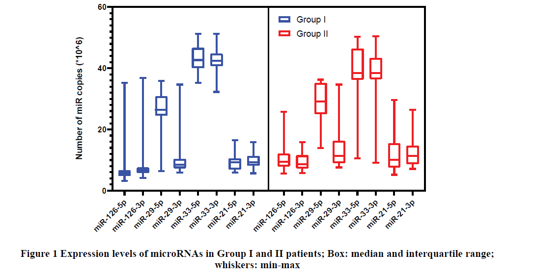
The analysis of miRs expression depending on atherosclerosis severity revealed a significant difference in miRs levels according to U-criteria (Table 3 and Figure 1).
| microRNA | Group I (ICA stenosis ≥ 50%) (*106 copies) | Group II (ICA stenosis <50%) (*106 copies) | p-value |
|---|---|---|---|
| miR-126-5p | 5.7 (4.8;6.62) | 9.4 (8.1; 11.8) | <0.001 |
| miR-126-3p | 6.64 (5.8; 7.52) | 8.7 (7.55; 11.45) | <0.001 |
| miR-29a-5p | 26.45 (24.6; 30.7) | 29.1 (25.5; 34.65) | 0.146 |
| miR-29a-3p | 8.46 (7.43; 11.4) | 11.4 (9.07; 15.79) | <0.001 |
| miR-33a-5p | 42.7 (40.5; 46.6) | 38.45 (36.3; 46.25) | 0.06 |
| miR-33a-3p | 42.45 (41.3; 44.6) | 38.4 (36.5;43.05) | 0.035 |
| miR-21-5p | 9.26 (6.98; 10.5) | 10.15 (7.78; 14.6) | 0.21 |
| miR-21-3p | 9.31 (8.24; 11.3) | 11.42 (8.72; 13.98) | 0.043 |
The expression levels of most microRNAs were statistically different between groups, with miR-126-5p/3p, miR-29- 3p, and miR-21-3p significantly lower in Group I, and miR-33a-3p-higher (Table 3, and Figure 1).
As seen from the Box Plot analysis most mature microRNAs which originate from opposite arms of the same premiRNA (denoted-5p and-3p) have roughly similar levels, except for the miR-29a-the expression of the -5p variant was three-fold higher than of its counterpart.
Routine blood tests did not show any relevant differences between the two groups (Table 4).
| Characteristics | ICA stenosis 50% or greater (n=30) (I) | ICA stenosis < 50% (n=20) (II) | p-value |
|---|---|---|---|
| Total cholesterol, mmol/l | 4.85 (4.2; 6.3) | 6 (4.5; 7.6) | 0.082 |
| Triglycerides, mmol/l | 1.38 (0.99; 2.04) | 1.89 (1.15; 1.99) | 0.534 |
| HDL, mmol/l | 1.76 (1.41; 2.28) | 1.8 (1.68; 2.21) | 0.85 |
| LDL, mmol/l | 1.99 (1.46; 2.74) | 2.5 (1.28; 2.9) | 0.645 |
| Fibrinogen, g/l | 3.44 (2.9; 4.17) | 3.8 (3.33; 4.26) | 0.232 |
| APTT, sec | 27.9 (26.5; 30.5) | 28.6 (25.1; 29.6) | 0.589 |
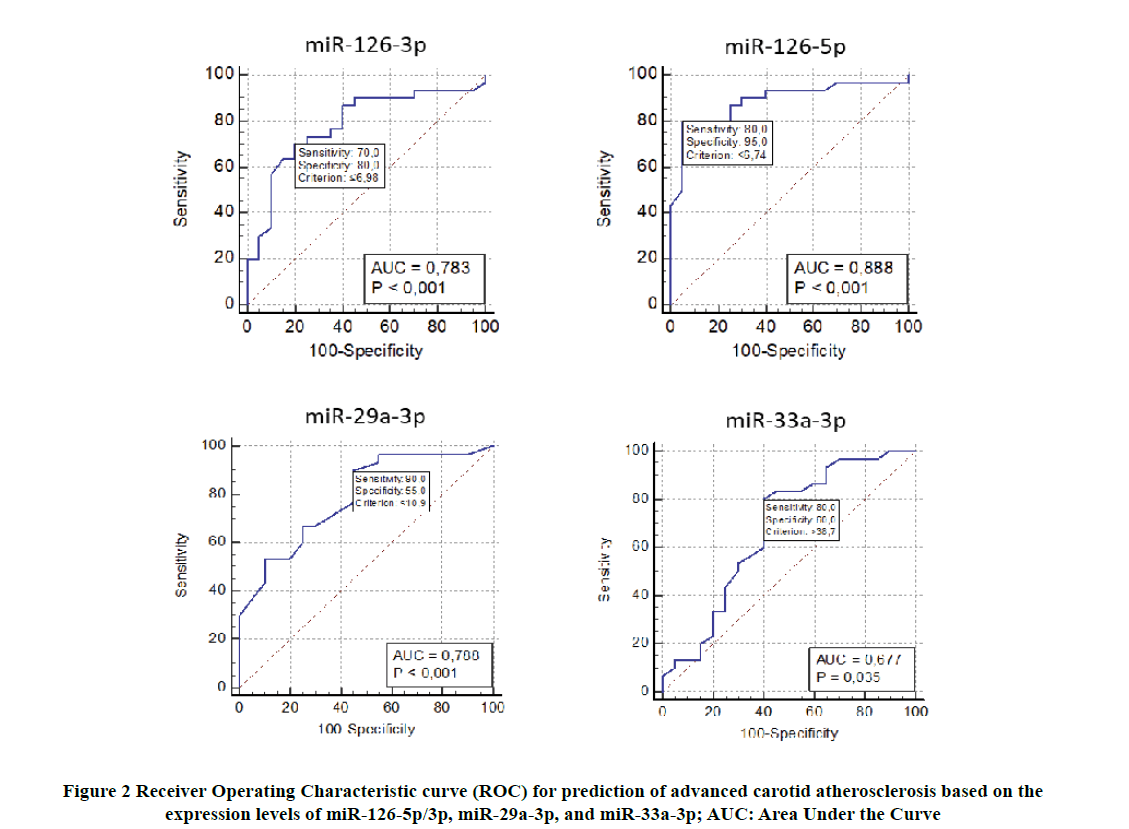
The predictive value of each parameter was assessed; an algorithm for assessing was identified. The model was checked by ROC analysis (Figure 2), the Area Under the Curve (AUC) was 0.89 for miR-126-5p (sensitivity 80%, specificity 95%, p<0.001), 0.78 for miR-126-3p (sensitivity 70%, specificity 80%, p<0.001), 0.79 for miR-29a-3p (sensitivity 90%, specificity 55%, p<0.001), 0.68 for miR-33a-3p (sensitivity 80%, specificity 60%, p=0.035).
Discussion
Carotid AS is one of the major risk factors for ischemic stroke with high levels of disability and mortality. There have been numerous studies that investigated the underlying pathological mechanisms of AS, but the molecular mechanisms and possibilities of precise biomarker diagnostics are not fully applied in clinical practice. MicroRNAs are an evolving biomarker of many physiological and pathological conditions, including atherosclerosis. A direct comparison of data from our study has shown that patients with carotid AS have a different profile of miR expression than healthy controls. The results of our study demonstrate that the expression of some miRs (in particular, miR-126- 5p/3p, miR-29a-3p, miR-21-3p) was remarkably down-regulated in patients with more severe carotid AS with only one miRNA (miR-33a-3p) up-regulated.
miR-126 is an endothelial-cell-specific miRNA encoded in the intron of epidermal-growth-factor-like domain 7 that gives rise to a pair of mature miRNAs, miR-126-3p and miR-126-5p. miR-126 is selectively expressed in endothelial cells, in embryonic stem cells-derived progenitor cells, and released by adipose-derived stem cells. miR-126 showed an important role in angiogenesis and vascular inflammation regulation moreover, it is suggested that miR-126 may be critical for the development and growth of organisms as targeted deletion of miR-126 in mice causes’ partial embryonic lethality [9]. miR-126 negatively regulates Vascular Cell Adhesion Molecule 1 (VCAM-1), which is required for the adhesion of leukocytes to the endothelium to initiate the recruitment of factors for inflammation [10]. Lower expression of miR-126 has been found in mice with carotid atherosclerotic plaque, while its target gene vascular cell adhesion molecule-1 was significantly up-regulated, which accelerated the progression of AS [11]. Moreover, it was found out that miR-126 reduces cytokine release and also decreases the progression of AS. miR-126-5p expression is essential for the replicative response of the endothelial cell to injury, like oxidative stress, by targeting the negative regulator of endothelial cell proliferation. Inadequate endothelium proliferation promotes lesion formation, which can be rescued by administration of miR-126 [12]. Zernecke, et al. found that the intravenous injection of miR-126 can inhibit AS in mice and reduce the formation of atherosclerotic plaques [13]. The pro-angiogenic impact of miR-126 was demonstrated by in vivo studies showing induction of blood vessel formation in the infarction region of the heart in a rat model of acute myocardial infarction [14]. miR-126 was found to be involved in the Mitogen-Associated Protein Kinase (MAPK) signaling pathway [15,16]. Our results seem to be corroborated by previous studies-both mature miR-126 (-5p and -3p) were expressed significantly lower in more prominent carotid AS patients. miR-126- 5p also had the most predictive value of all with 80% specificity and 95% sensitivity (AUC=0.888, p<0.001). These findings suggest atheroprotective functions of both miR-126 strands through different mechanisms of endothelial regeneration.
The miR-29 family, modulating mRNA level of collagen, inflammatory reaction, and other extracellular matrix genes, play a multifaceted role in tissue remodeling and vessel injury [7]. It is known that miR-29s are more highly expressed in Vascular Smooth Muscle Cells (VSMCs) than in other cell types and regulate VSMC functions, which are bound up with AS because VSMC proliferation is a major event of the AS [17]. miR-29a-3p shows a remarkable inhibitory effect on the expression of TNFa-induced adhesion molecules (VCAM-1, ICAM-1, and E-selectin) in vascular endothelial cells [18,19]. Some studies show that chronic miR‐29 treatment in a well‐accepted mouse model of AS increases indices of plaque stability, indicating a potential role for modulation of miR‐29 to affect plaque size and composition [20]. The miRNA-29 family has been extensively studied in various pathologies, including hepatic fibrosis, cardiac fibrosis, renal fibrosis, and pulmonary fibrosis. Recently, miR-29a has gained attention as a marker for progressive cardiovascular disease, in particular-endothelial dysfunction. miR-29a-3p can ameliorate TNFα-induced endothelial dysfunction by targeting tumor necrosis factor receptor 1, which, the authors of this study suggest, leads to a conclusion that miR-29a may be a potential novel target for the early prevention of atherosclerosis [21]. Our findings are in line with these data, but statistically significant differences between groups were identified only for the miR- 29a-3p variant. This is the more interesting, since its pair, miR-29a-5, was expressed nearly three-fold higher than that of its counterpart. Based on our data miR-29a-3p down-regulation can potentially be a marker in advanced carotid AS.
According to our data miR-21-3p expression was also lower in the first group. miR-21 has been suggested to regulate and promote vascular smooth muscle cell proliferation, but its role in atherosclerosis remains to be determined [22]. Yet, Wang, et al. could show prevention of in-stent restenosis by using an anti-miR-21 eluting stent in a humanized rat model [23]. Canfrán-Duque, et al. elucidated that the absence of miR-21 in macrophages results in an accelerated progression of AS, intra-plaque necrosis, and overall vascular inflammation [24]. Lack of miR-21 leads to impaired smooth muscle cell proliferation rates and enhanced apoptosis, leading to impaired vascular remodeling [25]. miR- 21 has been reported as an important mediator in human hypertension, abdominal aortic aneurysm development and expansion, atherogenesis, and myocardial infarction [26]. miR-21 is described as a “mechano-miR”, responding to arterial shear stresses. Some studies have demonstrated that miR-21 might be involved in the early stages of AS in hypertensive patients and is correlated with decreased levels of nitric oxide and endothelial nitric oxide synthase [24,27].
The only miR to be up-regulated in our study was miR-33a-3p-a miR involved in lipid metabolism. It was demonstrated before that miR33 has been shown to target multiple genes involved in individual steps of cholesterol metabolism. It also has been reported that the antagonism of miR-33 in experimental animal models results in increasing serum HDL and decreasing cholesterol levels in peripheral tissues, providing atheroprotective action [28]. Fine regulation of miR-33a/b could be a promising new approach to preventing or treating cardiovascular diseases in the future. It was shown that the application of anti-miR-33 promoted increasing HDL-C through ABCA1 upregulation and led to regression of atherosclerotic plaque in LDLR-deficient mice [29]. Moreover, miR-33a-deficiency reduced the progression of AS in apoE-deficient mice. miR-33a regulates lipid homeostasis by targeting ATP-Binding Cassette transporter A (ABCA1) and Sterol-Regulatory Element-Binding Proteins (SREBP1). miR-33a played an important role in inhibiting cholesterol efflux in macrophages via suppression of ABCA1 expression. So miR-33a expression level can be a candidate biomarker for the early detection of AS [30].
Our study has several limitations, among which the most prominent is the small cohort size; potential for selection bias; the compromised scope of discussions. Nevertheless, a common miRNA profile for advanced carotid atherosclerosis has been identified, which may supplement existing screening opportunities and preventive measures in the setting of cerebrovascular disease.
Conclusion
Carotid atherosclerosis is a chronic progressive pathology which may lead to acute and chronic cerebrovascular disease, yet in itself, it is heterogenic, with different biomarker profile for various stages. In our study, we have identified promising biomarkers of advanced CA-a a certain group of microRNAs which are down-regulated (miR- 126-5p/3p, miR-29a-3p, miR-21-3p) and, thus, may serve as potentially anti-atherogenic, and up-regulated (miR-33a- 3p), which exhibit proatherogenic properties. Our results may be useful for supporting further investigation to select potentially useful biomarkers for clinical practice.
Declarations
Conflicts of Interest
The authors declared no potential conflicts of interest with respect to the research, authorship, and/or publication of this article.
Funding
This study was funded by Grant No МК-3978.2019.7 of the President of the Russian Federation (Ministry of Science and Higher Education of the Russian Federation).
References
- Wu, Meng-Yu, et al. "New insights into the role of inflammation in the pathogenesis of atherosclerosis." International Journal of Molecular Sciences, Vol. 18, No. 10, 2017, p. 2034.
- Varela, Nelson, et al. "The current state of MicroRNAs as restenosis biomarkers." Frontiers in Genetics, Vol. 10, 2020, p. 1247.
- Azad, Fatemeh Mirzadeh, et al. "Small molecules with big impacts on cardiovascular diseases." Biochemical Genetics, Vol. 58, No. 3, 2020, pp. 359-83.
- Pereira-da-Silva, Tiago, et al. "Circulating microRNA profiles in different arterial territories of stable atherosclerotic disease: A systematic review." American Journal of Cardiovascular Disease, Vol. 8, No. 1, 2018, pp. 1-13.
- Joh, Jin Hyun, and Sungsin Cho. "Cardiovascular risk of carotid atherosclerosis: Global consensus beyond societal guidelines." The Lancet Global Health, Vol. 8, No. 5, 2020, pp. e625-26.
- Giral, Hector, Adelheid Kratzer, and Ulf Landmesser. "MicroRNAs in lipid metabolism and atherosclerosis." Best Practice and Research Clinical Endocrinology and Metabolism, Vol. 30, No. 5, 2016, pp. 665-76.
- Bretschneider, Maria, et al. "Activated mineralocorticoid receptor regulates micro‐RNA‐29b in vascular smooth muscle cells." The FASEB Journal, Vol. 30, No. 4, 2016, pp. 1610-22.
- Raskurazhev, A. A., et al. "Micro-RNA in patients with carotid atherosclerosis." Human Physiology, Vol. 46, No. 8, 2020, pp. 880-85.
- Yan, Yi, et al. "Deletion of miR-126a promotes hepatic aging and inflammation in a mouse model of cholestasis." Molecular Therapy-Nucleic Acids, Vol. 16, 2019, pp. 494-504.
- La Sala, Lucia, Francesco Prattichizzo, and Antonio Ceriello. "The link between diabetes and atherosclerosis." European Journal of Preventive Cardiology, Vol. 26, No. 2_suppl, 2019, pp. 15-24.
- Pan, Xudong, et al. "Atorvastatin upregulates the expression of miR-126 in apolipoprotein E-knockout mice with carotid atherosclerotic plaque." Cellular and Molecular Neurobiology, Vol. 37, No. 1, 2017, pp. 29-36.
- Tang, Feng, and Tian-Lun Yang. "MicroRNA-126 alleviates endothelial cells injury in atherosclerosis by restoring autophagic flux via inhibiting of PI3K/Akt/mTOR pathway." Biochemical and Biophysical Research Communications, Vol. 495, No. 1, 2018, pp. 1482-89.
- Zernecke, Alma, et al. "Delivery of microRNA-126 by apoptotic bodies induces CXCL12-dependent vascular protection." Science Signaling, Vol. 2, No. 100, 2009, p. ra81.
- Moghiman, Toktam, et al. "Therapeutic angiogenesis with exosomal microRNAs: An effectual approach for the treatment of myocardial ischemia." Heart Failure Reviews, Vol. 26, No. 1, 2021, pp. 205-13.
- D’Ardes, D., et al. "From endothelium to lipids, through microRNAs and PCSK9: A fascinating travel across atherosclerosis." High Blood Pressure and Cardiovascular Prevention, Vol. 27, No. 1, 2020, pp. 1-8.
- Hao, X. Z., and H. M. Fan. "Identification of miRNAs as atherosclerosis biomarkers and functional role of miR-126 in atherosclerosis progression through MAPK signalling pathway." European Review for Medical and Pharmacological Sciences, Vol. 21, No. 11, 2017, pp. 2725-33.
- Zheng, Bin, et al. "Regulatory crosstalk between KLF5, miR-29a and Fbw7/CDC4 cooperatively promotes atherosclerotic development." Biochimica et Biophysica Acta (BBA)-Molecular Basis of Disease, Vol. 1864, No. 2, 2018, pp. 374-86.
- Liu, Cui-zhong, Qi Zhong, and Yu-qing Huang. "Elevated plasma miR-29a levels are associated with increased carotid intima-media thickness in atherosclerosis patients." The Tohoku Journal of Experimental Medicine, Vol. 241, No. 3, 2017, pp. 183-88.
- Ulrich, Victoria, et al. "Chronic miR‐29 antagonism promotes favorable plaque remodeling in atherosclerotic mice." EMBO Molecular Medicine, Vol. 8, No. 6, 2016, pp. 643-53.
- Huang, Yu-qing, et al. "The association of circulating MiR-29b and interleukin-6 with subclinical atherosclerosis." Cellular Physiology and Biochemistry, Vol. 44, No. 4, 2017, pp. 1537-44.
- Deng, Xinrui, et al. "MicroRNA-29a-3p reduces TNFα-induced endothelial dysfunction by targeting tumor necrosis factor receptor 1." Molecular Therapy-Nucleic Acids, Vol. 18, 2019, pp. 903-15.
- Ji, Ruirui, et al. "MicroRNA expression signature and antisense-mediated depletion reveal an essential role of MicroRNA in vascular neointimal lesion formation." Circulation Research, Vol. 100, No. 11, 2007, pp. 1579-88.
- Wang, Dong, et al. "Local microRNA modulation using a novel anti-miR-21–eluting stent effectively prevents experimental in-stent restenosis." Arteriosclerosis, Thrombosis, and Vascular Biology, Vol. 35, No. 9, 2015, pp. 1945-53.
- Canfrán‐Duque, Alberto, et al. "Macrophage deficiency of miR‐21 promotes apoptosis, plaque necrosis, and vascular inflammation during atherogenesis." EMBO Molecular Medicine, Vol. 9, No. 9, 2017, pp. 1244-62.
- Jin, Hong, et al. "Local delivery of miR-21 stabilizes fibrous caps in vulnerable atherosclerotic lesions." Molecular Therapy, Vol. 26, No. 4, 2018, pp. 1040-55.
- Raitoharju, Emma, et al. "miR-21, miR-210, miR-34a, and miR-146a/b are up-regulated in human atherosclerotic plaques in the Tampere Vascular Study." Atherosclerosis, Vol. 219, No. 1, 2011, pp. 211-17.
- Cengiz, Mahir, et al. "Circulating miR-21 and eNOS in subclinical atherosclerosis in patients with hypertension." Clinical and Experimental Hypertension, Vol. 37, No. 8, 2015, pp. 643-49.
- Marquart, Tyler J., et al. "miR-33 links SREBP-2 induction to repression of sterol transporters." Proceedings of the National Academy of Sciences, Vol. 107, No. 27, 2010, pp. 12228-32.
- Rayner, Katey J., et al. "Antagonism of miR-33 in mice promotes reverse cholesterol transport and regression of atherosclerosis." The Journal of Clinical Investigation, Vol. 121, No. 7, 2011, pp. 2921-31.
- Kim, Soo Hwan, et al. "Aberrant expression of plasma microRNA-33a in an atherosclerosis-risk group." Molecular Biology Reports, Vol. 44, No. 1, 2017, pp. 79-88.


