Research - International Journal of Medical Research & Health Sciences ( 2020) Volume 9, Issue 9
The Prevalence of Endodontic Instrument Separation among Dental Practitioners and Dental Students in Riyadh, Saudi Arabia: A Cross-Sectional Study
Abeer AL Rumyyan1,2,3, Hamad Alissa1, Hamoud Alkuraidis1, Mohammed Sager1, Sulaiman Alraffa1, Ibrahim Alhumud1, Ahmad Alkhodair1*, Asim Aloraini1, Nawaf Almustafa1 and Jaser Alghamdi12King Abdullah International Medical Research Centre, Riyadh, Saudi Arabia
3Ministry of National Guard-Health Affairs, Riyadh, Saudi Arabia
Ahmad Alkhodair, College of Dentistry, King Saud bin Abdulaziz University for Health Sciences, Riyadh, Saudi Arabia, Tel: +966 11 429 9999, Email: Alkhadair.a@hotmail.com
Received: 03-Aug-2020 Accepted Date: Sep 21, 2020 ; Published: 28-Sep-2020
Abstract
Aims: This study aimed to investigate the prevalence of instrument separation and its management in Riyadh City.
Methods and Material: A survey was distributed in hard and soft copy forms. Target subjects were Undergraduate students, Dental interns, General practitioners, Postgraduates Endodontic, Advanced general dentistry (AGD), Saudi board advanced restorative dentistry (SBARD), and Endodontists. The questionnaire contained three domains: demographic data, incidence of instrument separation, management and follow up of instruments separation. Data were statistically analysed, and the significance level was set at p<0.05.
Results: The study includes 455 subjects. Determined percentage of instrument separation with hand file was 56.1% rotary file was 43.9%, nickel titanium alloy was 49.8% and stainless-steel was 50.2%. Comparable percentage of instruments separation in molars was more than other teeth (52.4%). Conclusion: The prevalence of instrument separation during root canal treatment was very high. Students and dentist awareness regarding causes and management of instrument separation should be increased to ensure successful root canal treatment.
Keywords
Dental practitioners, Dental students, Endodontic mishap, File separation, Root number
Introduction
The goal of endodontic treatment is to eradicate microorganisms from the root canal system, due to the fact bacteria are the causative factor for the development of apical periodontitis [1,2]. A study showed that there is a high success rate after the elimination of microorganisms from the root canal system before obturation [1]. When these measures are taking into account the success rate demonstrates to be as high as 94% [3,4]. Other studies showed incomplete removal of bacteria can result in an uncertain outcome of the root canal treatment [1]. During endodontic treatment, endodontists can face a variety of complications, one of the most is intraradicular instrument separation [5,6]. Instrument separation has as well been revealed to diminish the success rate by up to 14% when contrasted with those in which there was no instrument separation [7]. In another study done by Yousuf et al., a sum of 1748 root canal treated teeth were evaluated, 16 (0.9%) had instrument separation [6]. A study done by Iqbal, et al. [8], 37 rotary Nickle Titanium (NiTi) was fractured during endodontic treatment. the number of uses increases the percentage of the file to break. The angle of the curvature has no impact on file separation. However, regarding the radius of the curvature as the radius decrease the incidence of file separation increase. Most of the files fractured in the apical third [8]. According to Iqbal, et al. [8], A total of 81 files separated, the majority were NiTi rotary instruments. They observed that the most common tooth type that has file separation was the mandibular molar. The apical third is the most common site where the file separation occurs due to curvature and small diameter of the canal [8]. Moreover, the chance of having file separation in the apical third is 33 times higher than the coronal third and 6 times higher compared to the middle third [8]. Several clinical studies and series of case reports have reported the removal of separated instruments using ultrasonic devices, instrument removal systems, and Masserann kits [9-11]. Furthermore, it is essential to frequently follow-up the cases in the event of any further complication. This assists in clinical and radiographic assessment when the worsening of periapical tissues is recognized, endodontic apical surgery and extraction should be considered as valid treatment options [12]. Most studies focused on instrument separation in general but did not identify the relationship between the level of education and instrument separation which lead to a big gap in the literature regarding instrument separation epidemiology in terms of demographics, pulp status, and good sample size [8,13,14]. Shortage of studies with a good sample size that have been done in Saudi Arabia about file separation and have similar gaps in the literature. Therefore, this study aimed to assess the prevalence of instrument separation and its management in Riyadh City.
Methods
This is a cross-sectional observational study of the prevalence of instrument separation in Riyadh, Saudi Arabia. Ethical approval was obtained from the institutional review board committee at King Abdullah International Medical Research Centre, (RYD-IRB SP19/524/R) before the study. The sample size was calculated using an online sample size calculator with a margin of error of 5%, and a confidence interval of 0.95% for Riyadh population, the minimum recommended sample size for this study was 385 participants [15]. The study was conducted using a self-administered close-ended questionnaire. The questionnaire consisted of the following sections: first, demographic data including gender, professional status, type of sector, years of experience, and the number of cases per week. Second, the incidence of instrument separation and factors contributing to it. Third, treatment choice, management of mishaps, follow up, and prognosis. The questionnaire was taken from Pedir, et al. study [16] and was modified to meet the targeted population. The modification includes the professional status, number of cases per week, length of the separated instrument, and examination of the instrument during root canal treatment [16]. The data were collected by using both hard and electronic copy, and a consent form was provided on the first page of the questionnaire. The hard and soft copy was distributed by using a simple random sampling technique to five private and governmental universities, four governmental hospitals, and seventeen private clinics. Target subjects based on the inclusion criteria were all dental students in the 5th and 6th year since only 5th and 6th are allowed to operate on patients, dental interns, general practitioners, endodontic postgraduate, Advanced general dentistry (AGD), Saudi board advanced restorative dentistry (SBARD), and Endodontists. Fourth-year dental students and practitioners who do not perform root canal treatment routinely were excluded from this study.
Statistical Analysis
The data received were transferred in an excel sheet, then coded and analyzed using IBM statistical package for the social sciences software, version 22.0 (IBM, Armonk, New York). Chi-square test was used to assess the relationship between instrument separation with gender, professional status, working sector, cause of breakage, canal anatomy, type of tooth, and location of the canal. All statistical tests were declared significant at a p-value of 5% (0.05) or less along with a confidence interval of 95%.
Results
A total of 455 samples was obtained from 413 hard copy surveys and 43 online surveys, 233 working in the governmental sector, while 222 in the private sector. The demographic data of the participants are presented in (Table 1).
| Demographic | Frequency | Percentage (%) | |
|---|---|---|---|
| Professional Status | D3* | 78 | 17.10% |
| D4* | 113 | 24.80% | |
| intern | 102 | 22.40% | |
| General practitioners | 128 | 28.10% | |
| Endodontic postgraduate | 14 | 3.10% | |
| SBARD | 1 | 0.20% | |
| AGD | 9 | 2% | |
| Endodontist | 10 | 2.20% | |
| Working Sector | Private Sector | 222 | 48.80% |
| Government Sector | 233 | 51.20% | |
| Gender | Male | 348 | 76.50% |
| Female | 107 | 23.50% | |
*D3 and D4 are the 5th and 6th years of dental school respectively
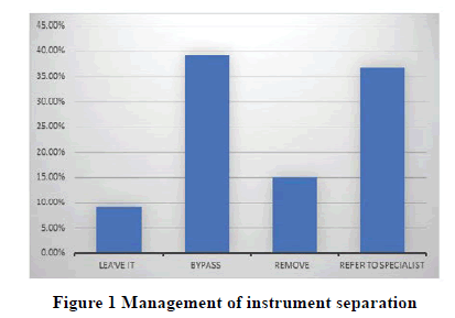
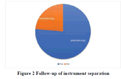
In the sample, a relation was found between the level of education and instrument separation of p=0.000, the percentage of instrument separation was 100% (n=14) of Endodontic postgraduates, 90% (n=9) of Endodontists, 82.8% (n=106) of General practitioners and 17.7% (n=20) of sixth-year students. An association was found between the type of file and instrument separation of p=0.000, the percentage of instrument separation was 56.1% (n=179) with hand file, 43.9% (n=140) with rotary file, 49.8% (n=158). Moreover, type of alloy appeared to play an important role in instrument separation of p=0.015, 49.8% (n=158) with NiTi alloy file, 50.2% (n=159) with stainless-steel alloy file. Another factor was type of tooth pf p=0.002, percentage of instruments separation in Molars was 52.4% (n=150), Premolars was 38.8% (n=111), Canines was 7% (n=20), and Incisors was 1.7% (n=5). It was found that the management of separated instruments and follow-up as presented in Figures 1 and 2 respectively. Other factors showed no significant relation with instrument separation.
Discussion
A significant association was found between the level of education and instrument separation as the higher the endodontic specialization the higher is the instrument separation, which can be explained by the higher number of endodontic cases performed per week, which is in agreement with a study conducted by Madarati, et al. [12]. on the contrary, Pedir, et al. found that general practitioners had the highest prevalence in instrument separation [16]. No previous studies reported an association between dental students and instrument separation. Also, the type of file plays an important role in instrument separation. It was found that rotary file separation was high when compared to hand files. This is due to the low yield and tensile strength of rotary compared to hand files resulted in an increased susceptibility to fracture at lower loads [17]. Similar findings were reported by Triantafyllia, et al. [18]. Furthermore, in this study the stainless-steel file separation was higher than NiTi. This is due to that NiTi files have more elastic, flexible, and fracture resistance than stainless-steel files [19,20]. When linking instrument separation and type of tooth, molar teeth found to have a higher prevalence of instrument separation than premolars and anteriors, this corresponds with previous studies [21-25]. This could be explained by Martin, et al. who demonstrated that the fracture rate could be impacted by the operator and the complexity of the canal anatomy [26]. In the present study, it was found that the preferred method for managing instrument separation was bypassing the separated file, given that bypass is the safest method because it involves removing a minimal amount of dentinal walls and is believed to contribute to a successful treatment outcome [27-29]. Also, it was found that most of the participants decided to follow-up their cases. Some investigations recommend following up the cases when removal or bypassing the separated instrument is impossible, high risk of mishap, or when instrumentation and obturation are coronal to the separated instrument [27]. The total number of respondents to the questions were different, that suggests the questions were freely answered by the participants. This study highlights the prevalence of instrument separation and will help to raise awareness among practitioners and improve the quality of endodontic treatment. Furthermore, it demonstrated the prevalence of instrument separation among dental students and dental practitioners were comparable to previous studies except in one aspect: the frequency of instrument separation concerning the level of education. This emphasizes the importance of further implementation of courses and a variety of educational methods concerning instrument separation. Limitations of this study were uneven number between different professional status and genders. Also, the present study did not include endodontic instrument separation prevention. Also, it suggests future researches to be conducted in a larger population since this study was limited to Riyadh City.
Conclusion
The prevalence of instrument separation during root canal treatment was very high. Students and dentist awareness regarding causes and management of instrument separation should be increased to ensure successful root canal treatment.
Declarations
Acknowledgement
The authors all the participated governmental and private clinics and universities in the research for their collaboration.
Funding
This research did not receive any specific grant from funding agencies in the public, commercial, or not-for-profit sectors.
Conflicts of Interest
The authors declared no potential conflicts of interest with respect to the research, authorship, and/or publication of this article.
References
- Sjogren, U., et al. "Influence of infection at the time of root filling on the outcome of endodontic treatment of teeth with apical periodontitis." International Endodontic Journal, Vol. 30, No. 5, 1997, pp. 297-306.
- Moller, AKE JR, et al. "Influence on periapical tissues of indigenous oral bacteria and necrotic pulp tissue in monkeys." European Journal of Oral Sciences, Vol. 89, No. 6, 1981, pp. 475-84.
- Imura, Noboru, et al. "The outcome of endodontic treatment: A retrospective study of 2000 cases performed by a specialist." Journal of Endodontics, Vol. 33, No. 11, 2007, pp. 1278-82.
- Lazarski, Michael P., et al. "Epidemiological evaluation of the outcomes of nonsurgical root canal treatment in a large cohort of insured dental patients." Journal of Endodontics, Vol. 27, No. 12, 2001, pp. 791-96.
- Al-Zahrani, Mohammad S., and Saad Al-Nazhan. "Retrieval of separated instruments using a combined method with a modified vista dental tip." Saudi Endodontic Journal, Vol. 2, No. 1, 2012, p. 41.
- Avoaka-Boni, Marie-Chantal, et al. "Frequency of complications during endodontic treatment: A survey among dentists of the town of Abidjan." Saudi Endodontic Journal, Vol. 10, No. 1, 2020, pp. 45-50.
- Yousuf, Waqas, Moiz Khan, and Hasan Mehdi. "Endodontic procedural errors: frequency, type of error, and the most frequently treated tooth." International Journal of Dentistry, 2015, pp. 1-7.
- Iqbal, Mian K., Meetu R. Kohli, and Jessica S. Kim. "A retrospective clinical study of incidence of root canal instrument separation in an endodontics graduate program: A PennEndo database study." Journal of Endodontics, Vol. 32, No. 11, 2006, pp. 1048-52.
- Crump, Merwyn C., and Eugene Natkin. "Relationship of broken root canal instruments to endodontic case prognosis: A clinical investigation." The Journal of the American Dental Association, Vol. 80, No. 6, 1970, pp. 1341-47.
- Hulsmann, Michael. "Methods for removing metal obstructions from the root canal." Dental Traumatology, Vol. 9, No. 6, 1993, pp. 223-37.
- Alomairy, Khalid H. "Evaluating two techniques on removal of fractured rotary nickel-titanium endodontic instruments from root canals: an in vitro study." Journal of Endodontics, Vol. 35, No. 4, 2009, pp. 559-62.
- Madarati, A. A., D. C. Watts, and A. J. E. Qualtrough. "Opinions and attitudes of endodontists and general dental practitioners in the UK towards the intra‐canal fracture of endodontic instruments. Part 2." International Endodontic Journal, Vol. 41, No. 12, 2008, pp. 1079-87.
- Almanei, Kholod Khalil. "Quality of root canal treatment of molar teeth provided by Saudi dental students using hand and rotary preparation techniques: Pilot study." Saudi Endodontic Journal, Vol. 8, No. 1, 2018, pp. 1-6.
- Tzanetakis, Giorgos N., et al. "Prevalence and management of instrument fracture in the postgraduate endodontic program at the Dental School of Athens: A five-year retrospective clinical study." Journal of Endodontics, Vol. 34, No. 6, 2008, pp. 675-78.
- Sample Size Calculator. https://www.calculator.net/sample-size-calculator.html
- Pedir, Samah Samir, et al. "Evaluation of the Factors and Treatment Options of Separated Endodontic Files among dentists and undergraduate students in Riyadh area." Journal of Clinical and Diagnostic Research: JCDR, Vol. 10, No. 3, 2016, pp. 18-23.
- Kj, Anusavice. "Phillips’ science of dental materials." St. Louis: WB Saunders, 2003.
- Vouzara, Triantafyllia, Maryam el Chares, and Kleoniki Lyroudia. "Separated instrument in endodontics: Frequency, treatment and prognosis." Balkan Journal of Dental Medicine, Vol. 22, No. 3, 2018, pp. 123-32.
- Walia, Harmeet, William A. Brantley, and Harold Gerstein. "An initial investigation of the bending and torsional properties of Nitinol root canal files." Journal of Endodontics, Vol. 14, No. 7, 1988, pp. 346-51.
- Bergmans, Lars, et al. "Mechanical root canal preparation with NiTi rotary instruments: rationale, performance and safety." American Journal of Dentistry, Vol. 14, No. 5, 2001, pp. 324-33.
- Wang, Nan-Nan, et al. "Analysis of Mtwo rotary instrument separation during endodontic therapy: A retrospective clinical study." Cell Biochemistry and Biophysics, Vol. 70, No. 2, 2014, pp. 1091-5.
- Ungerechts, C., Asgeir Bårdsen, and Inge Fristad. "Instrument fracture in root canals‐where, why, when and what? A study from a student clinic." International Endodontic Journal, Vol. 47, No. 2, 2014, pp. 183-90.
- Di Fiore, P. M., et al. "Nickel-titanium rotary instrument fracture: A clinical practice assessment." International Endodontic Journal, Vol. 39, No. 9, 2006, pp. 700-8.
- Cujé, J., C. Bargholz, and M. Hülsmann. "The outcome of retained instrument removal in a specialist practice." International Endodontic Journal, Vol. 43, No. 7, 2010, pp. 545-54.
- Al-Nazhan, Saad, Mustafa Hasan Al-Attas, and Nassr Al-Maflehi. "Retrieval outcome of separated endodontic instruments by Saudi endodontic board residents: A Clinical retrospective study." Saudi Endodontic Journal, Vol. 8, No. 2, 2018, p. 77.
- Martin, B., et al. "Factors influencing the fracture of nickel-titanium rotary instruments." International Endodontic Journal, Vol. 36, No. 4, 2003, pp. 262-66.
- Madarati, Ahmad A., Mark J. Hunter, and Paul MH Dummer. "Management of intracanal separated instruments." Journal of Endodontics, Vol. 39, No. 5, 2013, pp. 569-81.
- Al‐Fouzan, K. S. "Incidence of rotary ProFile instrument fracture and the potential for bypassing in vivo." International Endodontic Journal, Vol. 36, No. 12, 2003, pp. 864-7.
- Hulsmann, M., and I. Schinkel. "Influence of several factors on the success or failure of removal of fractured instruments from the root canal." Dental Traumatology, Vol. 15, No. 6, 1999, pp. 252-8.


